In contrast, na2/na2 stem has a "normal" stem appearance but the internode length is significantly shorter (c). The cross-sectional views of na2/na2 stem (i and j) reveal similar nodal and inter-nodal vascular arrangement, as found in the wild type (d and e). The elongation of tassel internodes occurs in na2/na2 (c).
Figures
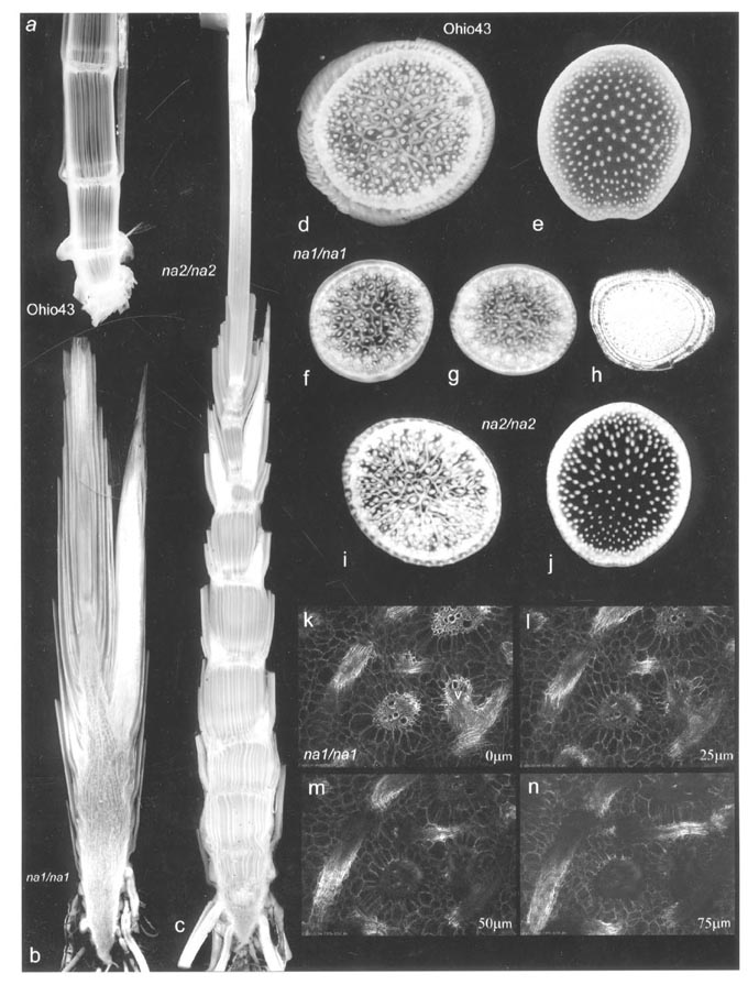
(previous page): (a) longitudinal section of a wild-type maize stem (Ohio43
inbred). (b) na1/na1 stem; (c) na2/na2 stem; (d and e) cross-sections
at node (d) and internodes (e) from wild-type plant; (f, g and h) cross-sections
at various levels of na1/na1 stem; (f) and (g) are physical sections
while (h) is an MRI section; (i and j) cross-section of na2/na2
stem at node (i) and internodes (j). All the plants used in this study
were grown at the field station of the University of Western Ontario, London,
Canada in the summer of 2000. The specimens were fixed in methanol, serial
sectioned with a razor blade using a specially made jig. The image was
obtained by using a modified Acer 600CU flat-bed scanner (600dpi optical
resolution, equipped with back-lighting) in liquid. (k-n) show a set of
optical sections (cross-sections) from na1/na1 stem. The optical
sections were obtained at various depths (0mm
� 75mm)
by two-photon florescence microscopy using 870nm near IR illumination.
An Olympus Fluorview FL300 confocal microscope equipped with a Spectra-Physics
Mai-Tai tunable Ti-sapphire laser was used for this study.
Return to the MNL 75 On-Line Index
Return to the Maize Newsletter Index
Return to the MaizeGDB Homepage