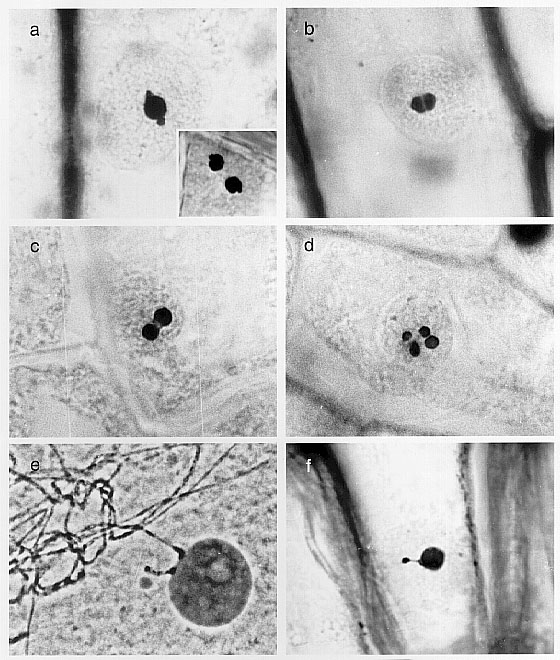
Our earlier study (Maillet et al. MNL 72:56-58,1998) on nucleoli has been extended to include an assessment of changes induced by specific environmental insults. There are several reports which describe changes in nucleoli during heat shock in terms of the loss of structure or the redistribution of specific proteins. Observation of intact silver stained nucleoli within epidermal cells of coleoptiles that were heat shocked 2, 4, or 6 hours (shifted from 27 C to 42 C) revealed a characteristic series of changes in nucleolar morphology. Although there is considerable variation in the response of cells from different regions of the tissue, at each time point most of the nucleoli in an area of the tissue would be at approximately the same stage. This variability may be the result of the mechanisms that control the heat shock response. Cells must receive a signal, either internal or external, to initiate transcription and translation, some heat shock proteins must move to the location where they act, and it is unlikely that all cells are equally responsive to heat shock. After two hours of HS the nucleoli had developed protrusions on their surfaces. The number of these structures corresponded to the number of NORs present in the genome. Cells from a diploid cultivar of maize (Ohio 43) had one or two protrusions. Since the nucleoli are viewed from one angle, the protrusions may not always be visible. Figure 1 shows heat shocked (two hours) Ohio 43 nuclei with one (a) and two (a insert) nucleoli. After four hours the many nucleoli had swollen and a furrow was present often in the middle of the nucleolus (Figure 1b). Often when two protrusions are observed in a diploid heat shocked cell they are at opposite ends of the nucleolus. Six hours after the initiation of HS many of the nucleoli appear to have been divided into two to four masses (Figures 1c and d).
In order to test the hypothesis that the protrusions are the sites where the NORs are attached to nucleoli, heat shocked coleoptile cells were examined in a cultivar that has additional NORs. The cultivar 2NOR has two NORs that are visible during early pachytene (Figure 1e), thus it would be expected that one to four nucleoli could be present (see the companion article for the number of nucleoli) in coleoptile cells, and that there would be one to four protrusions present after two hours of heat shock. Coleoptile cells that were heat shocked for two hours had one to four protrusions present on or near the surface of the nucleoli as expected, indicating that the number of protrusions are related to the number of NORs. In a very few nuclei a chromatin fiber could be seen connecting the micronucleolus to one of the protrusions on the nucleolus (Figure 1f).
These observations indicate that NORs can be made more distinct by a two hour heat shock and that the changes in nucleolar morphology appear to follow a characteristic series of stages.
Figure
1. Stages of changes in nucleolar morphology (a to d), early pachytene
in the 2 NOR cultivar (e), and a micronucleolus attached to a nucleolus
in the cultivar 2 NOR (f).
Return to the MNL 73 On-Line Index
Return to the Maize Newsletter Index
Return to the MaizeGDB Homepage