Isolated meiocyte culture of polymitotic microspores allows
direct observation of abnormal cell division
--Wolfe, KW, Liu, Q
Direct observation of meiotic divisions of developing maize microspores has become possible using the recently developed ADLP media (developed by Annette Chang, University of California Berkeley) for meiocyte culture. For the purpose of this study, efforts were concentrated on the development of techniques that allow the direct observation of po post-meiotic divisions, to yield information on the process of its post-meiotic abnormal cell cycles. Previously, no development beyond the tetrad stage has occurred in meiotically dividing cultured microspores. Here however, we report that during isolated po meiocyte culture, development proceeds through the tetrad stage and also through a number of subsequent cell cycles. These methods and results may be useful to the study of cell cycle regulation and the process of cell division.
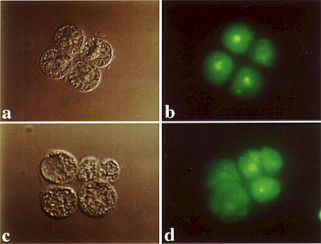
Meiocytes and microspores were isolated and cultured in ADLP media. To visualize chromatin structure, some cultures included SYTO 12 Live Cell Nucleic Acid Dye (Molecular Probes, Eugene OR) and observed for a period up to 1 week. After the second meiotic division, microspores remained in the tetrad stage (Figure 1a) a short time before chromatin condensation (Figure 1b) and mutational cell division occurred (Figures 1c and 1d). The best results were obtained with cells isolated immediately following meiosis II. Cells isolated both before and during the meiotic divisions were observed to continue development through both meiotic divisions and post-meiotic divisions, although to a much lesser degree. Meiotic divisions were noted to be synchronous, in contrast to the post-meiotic divisions, which were very asynchronous.
The timing of extra cell divisions varied widely and frequently, as cells within the same tetrad were observed to divide in a very non-synchronous manner. Often one cell of a tetrad would undergo many extra cell divisions while another in the same tetrad would undergo only one or two. Chromosome condensation was always observed in cells cultured with the SYTO 12 living cell nucleic acid fluorescent dye following the second meiotic division, preceding post-meiotic divisions. Cells incubated with SYTO 12 were not observed to go through the same extent of extra cell divisions, and the timing of extra cell divisions was noticeably longer.
The culture conditions do not alter microspore development appreciably from what is witnessed during in vivo microsporogenesis, although more work is underway for a complete characterization of cell division timing and behavior. Results obtained so far indicate that cells proceed through the mutational cell cycles to the same extent, and the timing of the divisions appears to match those observed in vivo, without the influence of the surrounding somatic anther tissue.
Figure 1. Isolated culture of po tetrads. a) a po microspore before post-meiotic divisions; b) chromatin is observed to condense before cells divide; c) the same cell pictures in (a), after the first post-meiotic division; d) chromatin is observable throughout post-meiotic divisions.
Return to the MNL 72 On-Line Index
Return to the Maize Newsletter Index
Return to the MaizeGDB Homepage