A confocal laser scanning microscopy study of 4 embryo specific
mutants
--Elster, R, Bommert, P, Werr, W
Embryo specific (emb) mutants in maize are specifically blocked in embryogenesis whereas endosperm development - unlike in defective kernel mutants - is not obviously affected. Phenotypic characterization in a large collection of 51 emb mutants was accomplished, so far, either in fully mature (40 to 60 days after pollination (DAP)) and fresh dissected kernels or in rehydrated seeds. A wide range of promising developmental blocks, interfering with early and late stages in embryo development, could be identified (Sheridan and Clark, Plant J. 3:347-358, 1993; Clark and Sheridan, Plant Cell 3:935-951).
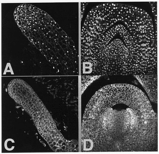
In order to have a more detailed view of the aberrant mutant embryo morphology, we have analyzed a subset of this mutant collection by use of a confocal laser scanning microscope (CLSM). Mutant embryos from four different lines, emb5 (former emb*-8518), emb9 (former emb*-8521), emb*-8537, and emb*-8542, harvested between 8 DAP and 20 DAP, were fixed, stained with propidium iodide and analyzed according to Running et al. (Methods in Cell Biol. 49:217-229, 1995). Compared to their wild type siblings all 4 mutants show retarded development already at 10 DAP and are easily distinguishable at this stage. However, at 15 DAP the specific blocks become more evident: emb5 and emb9 are blocked at a late proembryo/ early transition stage (see Figure 1) and fail to establish a radial asymmetry as wild type siblings do by induction of a shoot meristem (SM) and a scutellum. Mutants of this type are unable to overcome the block and even at later stages in kernel development the embryos still reside in a late proembryo or early transition stage. Cells of emb5 and emb9 mutant embryos are frequently highly vacuolized, indicating a terminal differentiation.
In contrast, mutant embryos from lines emb*-8537 and emb*-8542 show induction of a scutellum and organization of a region of cells resembling a SM are visible. At least for emb*-8537 we never found evidence for the formation of a coleoptile or embryonic leaves. Some emb*-8542 embryos analyzed at later stages, however, showed hyperplasia-like outgrowth and further embryonic tissues were formed, leading to structures which lack major plant organs, as roots or shoots are not produced.
Presently we are in the process of refining our CLSM data with an in situ analysis of mutant embryos at different developmental stages after pollination. By use of specific markers, commonly associated with the existence of a SM, we will try to establish whether the formation and function of this meristem in our mutants is affected also in molecular terms.
Figure 1. 1A shows a homozygote emb5 embryo 15 DAP which is blocked in an early transition stage whereas wild type siblings are already at leaf stage 1 (seen in 1B). 1C and 1D show the corresponding pictures for emb9.
Return to the MNL 72 On-Line Index
Return to the Maize Newsletter Index
Return to the MaizeGDB Homepage