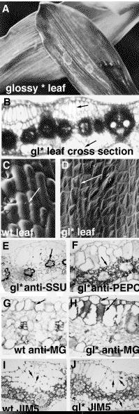
We also performed immunolocalization experiments (immunogold with silver enhancement, followed by counterstaining with basic fuschin) to determine the identity of the extra cells found in glossy* regions of the leaf. We used two different kinds of antibodies, antibodies to photosynthetic proteins and antibodies to cell wall compounds. Antibodies to Small Subunit of RuBisCo (SSU) bound to chloroplasts in the bundle sheath cells in both the wild type and glossy* sections (Fig. 1E). The extra cell regions in the glossy* leaf did not stain, indicating that these cells probably do not have photosynthetic activity or bundle sheath identity. Antibodies to Phosphoenol Pyruvate Carboxylase (PEPC) bound to the mesophyll cells in both wild type and glossy* sections (Fig. 1F). The extra cell regions did not stain, indicating that the extra cells are not active in C4 photosynthesis and do not have mesophyll identity. The anti-MG antibody recognizes mixed-linkage glycans in the cell wall of certain cells. Anti-MG stained the cell walls of the epidermal cells, midrib cells, and xylem in vascular bundles of both the wild type (Fig. 1G) and glossy* leaves (Fig. 1H). The bundle sheath and mesophyll cells did not show reactivity to the anti-MG antibody. The extra cells in glossy* leaves stained in their cell walls. This indicates that the cell walls of the extra cells are more like epidermal or midrib cells than bundle sheath or mesophyll cells, thus confirming the results from the photosynthetic antibodies. JIM5 is a monoclonal antibody against unesterified polygalacturonic acid and mostly decorates the corners where two epidermal cells join together. Therefore, we used this antibody to determine if the extra glossy* cells have epidermal identity. The corners of epidermal cells stained in both wild type (Fig. 1I) and glossy* leaf sections (Fig. 1J). In addition, the midrib cells also stained in the corners. The extra glossy* cells, however, did not show this same pattern of staining. Therefore, we can conclude that the extra cells in the glossy * mutation do not have bundle sheath, mesophyll or pure midrib cell identity. They are also not identical to epidermal cells.
Figure 1. A: Juvenile leaf of glossy* plant showing regions of abnormal leaf surfaces. B: Transverse section of gl* leaf showing regions of extra cell layers (arrows). C: SEM of a normal leaf surface showing cells with crenulated margins (arrows). D: SEM of gl* leaf surface showing abnormal cells with no crenulated margins (arrow). E: Transverse section of gl* leaf probed with anti-SSU antibody showing staining in the bundle sheath cells (arrow). F: Transverse section of gl* leaf probed with anti-PEPC antibody showing staining in the mesophyll cells (arrow). G: Transverse section of wild-type leaf probed with anti-MG antibody showing staining in the epidermal cells (arrow). H: Transverse section of gl* leaf probed with anti-MG antibody showing staining in the epidermal cells and extra cell layers (arrow). I: Transverse section of wild-type leaf probed with JIM5 antibody showing staining in the corners of epidermal and midrib cells (arrow). J: Transverse section of gl* leaf probed with JIM5 antibody showing staining in the epidermal cells and extra cell layers. However not all cell junctions are stained (arrows)
Return to the MNL 71 On-Line Index
Return to the Maize Newsletter Index
Return to the MaizeGDB Homepage