Ovary manipulation: transformation and in vitro maturation
--Ding, Q; Xie, Y; Dai, J; Mi, J; Kuo, Z
Ovary injection has been established as a transformation method (MNL 70:13-14) which has an obvious advantage: avoiding the limitation of genotypes on tissue culture and regeneration. On the other hand, this method has obvious shortcomings also: experience dependence and low efficiency. In order to improve this method some subjects were studied.
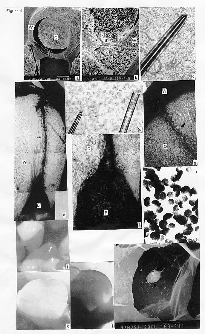
Carbon powders were used as tracers under the light microscope. Sampling at different times after injection, it was found that carbon powders could be injected into ovules and aggregate around the embryos along with their development. It was concluded that when the fertilized egg divided it could absorb nutritives from cells around itself and inert carbon particles dyed the embryo black (Figure 1e,f).
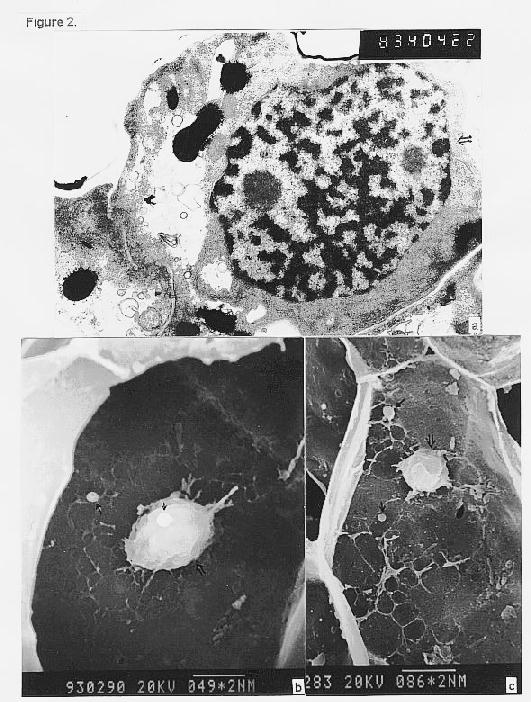
For detailed information, tungsten particles were used as tracers under electron microscope according to the procedure for preparation of DNA for bombardment. The result showed that tungsten particles could be found in embryo cells four days after injection (Figure 1i). In this stage the embryo cells have a large nucleus (Figure 2a), after sampling and treatment the nucleus sometimes could stick to cell wall and tungsten particles could stick to the cell wall too (Figure 2b,c). Interestingly, one tungsten particle adhered to a nucleus that had stuck to the cell wall (Figure 1i, Figure 2b). These results could provide a demonstration for the pathway of donor DNA delivered into egg and embryo cells.
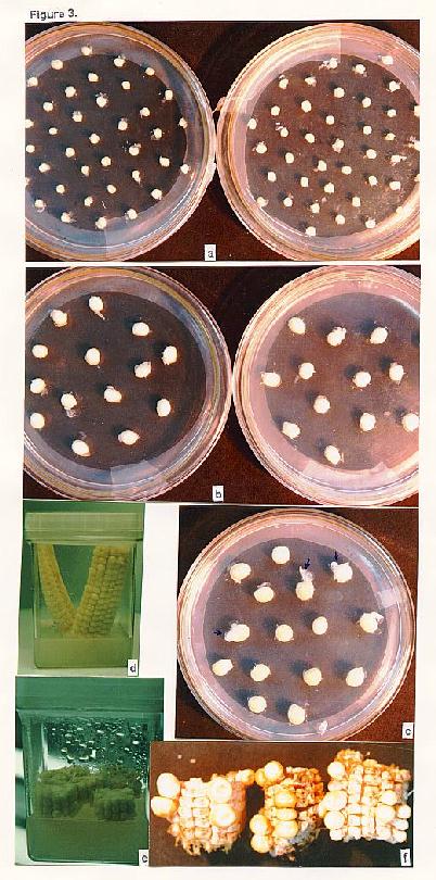
The main problem of ovary injection techniques has been that the injected ovaries were damaged by glass tubes and fungi, especially under the field condition. So I tried to culture isolated ovules and ovaries in order to increase the efficiency (Figure 1j,k,l; Figure 3). After two months culture the isolated ovules could just enlarge to 10 fold of their original size but with abnormal embryos. Although these ovules could be induced to calli they could not develop into embryos and plantlets. Isolated ovaries could develop into normal embryos but with very poor endosperm. Finally I cultured young fertilized ears. Normal seeds matured. After donor DNA injection, there are seeds matured but a lot of ovaries failed to produce seed (Figure 3f). At present about 180 injected seeds have been obtained and molecular identification is performed.
Figure 1. a: young ovary with ovule (O) and ovary wall (W); b: sac (S), ovule (O) and ovary wall (W) of ovary; c: tip of microcapillary (290X); d: comparison of tips of different microcapillaries (150X); e: carbon powder dyed embryo (E), normal ovule (O) and the trace (arrow) of injection (27X); f: carbon powder dyed embryo (E) (81X); g: trace of injection (arrow) (62X); h: tungsten particles (5000X); i: nucleus (double arrow) and tungsten particles (arrow) stuck to cell wall; j: isolated ovules (11X); k: growing ovule (13X) l: enlarged ovule (14X).
Figure 2. a: embryo cell with a large nucleus (4435X); b: detailed picture of nucleus (double arrow) and tungsten particles stuck to cell wall; c: nucleus (double arrow) and tungsten particles (arrow).
Figure 3. a: newly isolated ovaries; b: growing ovaries; c: some ovaries budding (arrow); d: cultured ears with vertical section; e: cultured ears with horizontal section; f: matured seeds after injection.
Return to the MNL 71 On-Line Index
Return to the Maize Newsletter Index
Return to the MaizeGDB Homepage