Agrobacterium-mediated gene transformation in maize
--Ishige, T
Generally, genetic transformation in grasses has been achieved by particle bombardment of intact tissues or electroporation of protoplasts. Recently Agrobacterium tumefaciens has been used to introduce foreign genes into rice chromosomes, and fertile rice transformants were obtained (Hiei et al., Breeding Science Suppl. 1:52, 1994). We report here the gene transformation of maize using the Ti-plasmid vector of A. tumefaciens.
Maize calli were initiated from immature embryos of F2 seed of an A188/B73 cross. Type II calli were selected and their regenerability was evaluated. The chimeric gene RB-NoS-NPTII-35S-HPT-35S-GUS-LB was constructed in PBI 101 binary vector and was transformed into LBA4404 strain of A. tumefaciens by electroporation. Maize calli were co-cultured with A. tumefaciens for three days in liquid N6 medium containing 2 mg/l of 2,4-D and the calli were transplanted in N6 selection media containing 2 mg/l of 2,4-D, 0.25 mg/l of hygromycin B, 3 mg/l of cefotaxime and 0.3% of gelrite. The GUS activity of selected calli was analyzed by staining the intact tissues. The calli were transferred to N6 regeneration medium lacking hormones and containing 0.25 mg/l of hygromycin B, 3 mg/l of cefotaxime and 0.3% of gelrite. The regenerated shoots were transplanted in soil and the genomic DNA of the leaf was extracted to confirm the integration of the introduced gene by PCR and Southern blot analysis.
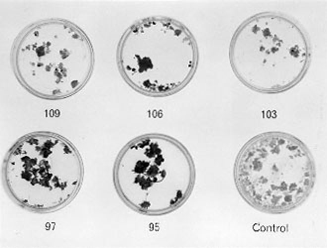
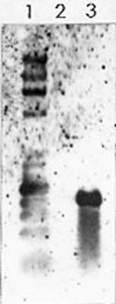
All of the callus lines infected with A. tumefaciens showed a blue color due to their GUS activity and GUS activity was not expressed in control calli in the absence of Agrobacterium infection (Fig. 1). The Southern blot analysis and PCR showed that the introduced gene was integrated in the corn genome (Fig. 2). A gene transfer method of maize using A. tumefaciens infection was thus developed.
Figure 1. Expression of GUS in maize tissues after co-cultivation with A. tumefaciens. The numbers are given to distinguish the calli from other cell lines of the F2 embryo. The GUS activity was determined by staining cells with X-gluc. The activity level varied among callus lines. Control calli without gene transformation did not show any GUS activity.
Figure 2. Southern analysis of the transformed maize. Lane 1: hHindIII, HaeIII size markers. 2: control. 3: transformed maize by A. tumefaciens. DNA (10µg) was digested with BamHI, and separated by electrophoresis in a 0.7% agarose gel. The DIG-labeled DNA of the HPT region was used as a probe of hybridization.
Return to the MNL 70 On-Line Index
Return to the Maize Newsletter Index
Return to the MaizeGDB Homepage