A method for studying vascular bundles in 3D
--Ping-chin Cheng
To study the arrangement of vascular bundles in a chunky specimen is a difficult task; frequently it relies on time consuming reconstruction of serial sections. Here we report a method which can be used for stereoscopic viewing of vascular bundles in bulky specimens (as large as a longitudinally bisected ear). The specimen preparation method described here can also be used for confocal microscopy.
The procedure involves fixing tissues in a 3:1 EtOH/acetic acid fixative. The tissue is left in the fixative until it is completely decolored. For a large piece of tissue, it may be necessary to make a few changes of fresh fixative. This process may take days to weeks. Then the tissue is degassed in an air-tight glass jar filled with gas-depleted water; degassed water is obtained by cooling down boiling water in a completely filled bottle capped with an air-tight cover. This degas step is essential for tissues which contain trapped air bubbles. For example, mature maize stem generally has a whitish appearance as a result of strong light scattering from the large number of air pockets. By immersing the air-containing tissue in a jar of degassed water, the trapped air can be slowly dissolved away, and finally the tissue is completely waterlogged. This process is best to be carried out in a closed jar which is topped to its rim with gas-depleted water in a 4C refrigerator. Although this degas process can be carried out at room temperature, the lower temperature enables a larger amount of gases to be dissolved in the bathing water, and hence is faster in removing the trapped air bubbles from the tissue. A small crystal of thymol should be added to the bathing water to prevent the possible growth of mold or bacteria.
When the tissue is completely free of air bubbles, the tissue is then soaked in a 0.5% (w/v) sodium metabisulfite solution. The length of this treatment ranges from an hour to several days depending on the size of the tissue. Then the tissue is transferred to a diluted Schiff reagent (prepared by adding 0.1g basic fuchsin to 85ml water with 1.9g sodium metabisulfite and 15ml 1N HCL; shaken for two hours and then decolorized with 200mg activated charcoal) at 4C, the staining time ranging from an hour to several days. It is important to use a jar with an air-tight cover to avoid the loss of SO2 from the solution. For large pieces of tissue, a standard canning jar works well. After staining, the tissue is differentiated in a sodium metabisulfite solution (0.5% w/v) for many changes. Complete removal of unbound dye at this step is essential to ensure the final preparation has the lowest background staining. Then the tissue is dehydrated through an EtOH series followed by several changes of absolute EtOH, 1:1 EtOH/methyl salicylate and finally cleared in several changes of pure methyl salicylate. To ensure a high degree of transparency in the final preparation, complete removal of tissue water by absolute EtOH is essential. The result is a highly transparent specimen with vascular bundles stained in purple.
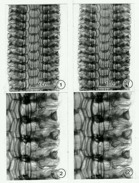
The stereogram (a stereo-image pair) is one of the most commonly used methods to present 3D structures. A stereogram can be obtained from a specimen by taking photographs of a specimen at two different viewing angles which simulate the appropriate parallax of the human eyes. On the other hand, a stereogram can be generated by a computer from a volumetric data set obtained by a laser scanning confocal microscope or tomographic system. The angle between the two images of a stereogram can be determined by a simple formula (Hudson and Makin, J. Phy. E. Sci. Inst., 3: 311, 1970). To photograph the specimen, a panchromatic film such as Kodak T-max 100 can be used. A green filter (Tiffen 58) will provide excellent contrast for a thin specimen (such as a leaf blade). However, for a bulky specimen (e.g. longitudinal section of an ear or a stem) where background staining is relatively high, the use of a red filter (Kodak Wratten 25 or Tiffen 25) is recommended. The specimen should be completely immersed in methyl salicylate in a flat-bottom glass container. An evenly illuminated light-box can be used as the light source. For microscopic tissues, confocal microscopy operated in epi-fluorescent mode using the 488nm Ar laser line as the excitation wavelength is ideal. A 550nm long-pass filter is used for the fluorescent detector. Serially cut optical sections obtained by the confocal microscope can then be used for generating the stereogram in a computer. For better stereo perception, it is important to avoid orienting fiber-like structures (such as vascular bundles in a stem) parallel to the base of the stereogram. Figure 1 shows a stereogram of a longitudinal slice of an ear inflorescence (var. Golden Beauty). Figure 2 is a higher magnification view showing vascular bundles associated with spikelets.
[Ed. note: With the original prints, I have been able to 'experience' a stereo image by crossed-eyes tripling and concentration on the middle image.]
In addition to simple stereoscopic viewing, the specimen preparation method mentioned above opens up possibilities of using optical tomography and multidimensional image analysis for true 3D reconstruction and interactive visualization of the vascular bundles on a computer. This work was supported by a grant from the National Science Council of the Republic of China (NSC 83-0211-B-001-001) during the author's sabbatical leave at the Institute of Botany, Academia Sinica, Taipei.
Return to the MNL 69 On-Line Index
Return to the Maize Newsletter Index
Return to the MaizeGDB Homepage