Determining the nuclear volume in a pollen grain by using laser scanning
confocal microscopy and multi-dimensional image analysis
--Ping-chin Cheng and J. K. Samarabandu
We have developed an automatic image processing tool for determining the volume of vegetative and sperm nuclei in a maize pollen grain. The procedure involved collection of confocal fluorescence images from a DAPI-stained/Feulgen-stained pollen grain at a 0.5 µm (or 1 µm) sectioning interval and processing the 3D image data with an automatic image processing system developed at our laboratory. To prepare for confocal microscopy, the stained pollen was dehydrated in EtOH, and cleared in methyl salicylate (Cheng et al., in Multi-dimensional Microscopy, P. C. Cheng et al., eds., Springer-Verlag, New York, pp. 339-380, 1993).
An attempt at manually contouring the cell nucleus in a sea urchin embryo in 3D was reported by Summers, RG et al. (J. Electron Micro. Tech. 18:24-30, 1990). Apart from being labor intensive, this 3D digitization technique suffers from the inaccuracies of manual 3D tracing related to the depth perception of the operator (Cheng et al., in Visualization in Multi-Slice Imaging Microscopies, A. Kriete, ed., VCH-Publ., Weinheim, pp. 361-398, 1993b). However, it does demonstrate that reducing a stack of confocal images to a 3D graphic representation helps to visualize and analyze complex tissue. This procedure also significantly reduces the computational burden in an interactive operation. These image analysis tools can also be employed for numerical and volumetric study of cell nuclei.
To overcome the disadvantage of manual tracing, an automatic data reduction procedure based on multi-dimensional image analysis algorithms was developed in our laboratory. We also developed a system to visualize and extract morphometrical parameters from the data generated by this method (Cheng et al., 1993; Samarabandu, in Multi-dimensional Microscopy, P. C. Cheng et al., eds., Springer-Verlag, New York, pp. 231-250, 1993). Our confocal image processing system is implemented as a set of tools whose activities are coordinated by a blackboard control structure and is modeled after the image understanding model introduced by Kanade, T (Comp. Graph. Imag. Proc. 13:279-297, 1980).
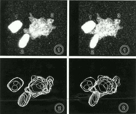
Figure 1 is a stereogram of a vegetative and two sperm nuclei stained
by Feulgen reaction. The image
was obtained by optical sectioning of the pollen grain at 1 µm
intervals with an Olympus GB-200 laser scanning confocal microscope. The
514nm green line of a 25mW Ar ion laser was used as the excitation wavelength,
and red fluorescence was recorded. Note the two intensely stained sperm
nuclei and a highly convoluted vegetative nucleus. Figure 2 shows the surface
contours of the nuclei generated by our automatic image processing program.
The program, at the present time, does show some difficulty in contouring
weakly stained fine projections of the vegetative nucleus. With the contour
generated, the volume of the nuclei can be calculated. In this case, the
volumes of the two sperm nuclei are (1) 249 µm3 and (2) 209 µm3;
the vegetative nucleus (3) is 973 µm3.
Figures
1 and 2. Stereogram and surface contours.
Return to the MNL 68 On-Line Index
Return to the Maize Newsletter Index
Return to the MaizeGDB Homepage