Recently, there has been some interest in light activated compounds, so-called phototoxins. We decided to extract maize leaves to see if any such compounds could be identified using a bacterial assay. After several trials we settled on extracting maize leaves with 70% methanol in a Waring blender going full speed for 2 min.
The first successful response came from extracts of Fr 632 Ht1 using the leaves of 5-6 leaf maize plants. The standard procedure has been to grind 10-20 grams of leaves without the sheath. The methanol mixture is 70% methanol, 30% water with one drop of beta-mercaptoethanol per 10 ml. The grinding ratio was 1 g of tissue per 10 ml of 70% methanol. After grinding, the mixture was spun in a Sorvall centrifuge for 10 min at 5,000 rpm. The liquid was taken off with a Pasteur pipette and spotted immediately onto filter discs, held by pins. The remaining liquid was stored at -20 C. Storage for several weeks did not seem to reduce activity.
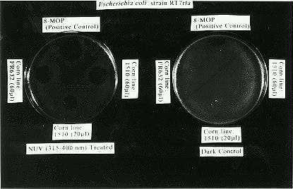
Cultures of Escherichia coli strain RT 7 rfa were grown the night before on Petri plates. Next morning the dry filter paper discs with the leaf extracts were allowed to absorb and diffuse on the Petri plates for 1 1/2 hours. A control compound, 8-MOP, was also spotted and blotted. Duplicate plates were always made up as dark controls. After the absorption from the discs was completed, and the paper removed, the plates were irradiated with UV-A (315-400 nm) for one hour. The other half of the plates (dark controls) were not. After the hour all of the plates were put at 37 C to grow until the next day, when they were examined. The UV-A 8-MOP control usually gave lethal circles, 25 mm in diameter. The positive genotypes with 0.060 ml of methanol extract had rings of killing nearly that large. Measurements of these diameters gave the bioassay a semi-quantitative measure. A typical experiment is shown in Figure 1.
The initial response was from a Ht1 genotype. Other maize stocks we tried were negative or had slight responses. To do a proper experiment we obtained a large series of inbreds that were Ht1 converted and ht1 from Illinois Foundation Seed. As seen in Table 1 below there was no correlation between Ht1 and reactivity.
Table 1.
| Genotype | Reactivity | Diameter of reactive zone (mm) |
| O7A ht1 | - | |
| O7A Ht1 | - | |
| M14 ht1 | +- | 6 |
| M14 Ht1 | - | |
| FR Mo17 rhm, ht1 | + | 9 |
| Mo17 Ht1 (Callahan) | - | |
| W22G ht1 | - | |
| W22G Ht1 | + | 10-11 |
| B37 ht1 | + | 9 |
| B37 Ht1 | +- | 3 |
| Oh51A ht1 | - | |
| Oh51A Ht1 | + | 8 |
| B68 ht1 | - | |
| B68, rhm, Ht1 | + | 16 |
| A619 ht1 | + | 9 |
| A619 rhm, Ht1 | - | |
| A635 ht1 | - | |
| A632 Ht1 | - | |
| MS1334 ht1 | - | |
| MS1334 Ht1 | - |
The most reactive genotype was a B68 inbred. B68 is known to have a high content of DIMBOA. For this reason we obtained seed from K. Simcox, K that are DIMBOA negative (bx/bx). In Table 2 bx/bx leaves are quite reactive in the bioassay. We are left without leads as to the role of this phototoxin in the plant. Finally, in mature plants (B37 ht1) we determined that the phototoxin was evenly distributed. The oldest leaves had the same reactivity as the youngest leaves on a per gram basis.
Table 2.
| Genotype | Reactivity | Diameter of reactive zone (mm) |
| bx/bx (Simcox) | + | 18 |
| Bx/Bx | +- | 3 |
| bx/bx (#16, Simcox) | + | 20 |
| B68 Ht1 | + | 16 |
| B68 ht1 | - | |
| B37 Ht1 | +- | 3 |
| B37 ht1 | + | 9 |
A number of varied experiments had been planned, including identifying the compound(s) by chemical methods. However the senior author (RWT) met an untimely death more than a year ago, so the studies were ended.
Figure
1. Petri dishes with E. coli cultures spotted with methanol
extracts of maize leaves. The plate on the left received UV, the right
did not. FR632 is an inbred from Illinois Foundation Seed. 1510 is a Mexican
flint. The clear circles are where the phototoxin killed the bacteria.
Return to the MNL 68 On-Line Index
Return to the Maize Newsletter Index
Return to the MaizeGDB Homepage