Anthocyanins extracted from plants absorb ultraviolet radiation. Measurements in situ demonstrate that the epidermis of plants absorbs 95%-99% of incoming UV-B radiation; flavonoid compounds (such as anthocyanins) and cuticular waxes are most likely the agents of UV absorption (Stapleton, Plant Cell 4:1353, 1992). Because anthocyanins and other flavonoids absorb radiation in the UV range, it is commonly suggested that these compounds could shield plant DNA from UV damage. This hypothesis has not previously been tested, however, nor has the extent of protection been quantified. In maize there are at least five anthocyanins present in purple tissues (Harborne and Gavazzi, Phytochem. 8:999, 1969; Styles and Ceska, Phytochem. 11:3019, 1972). We have investigated the ability of these compounds to protect DNA in situ from UV damage.
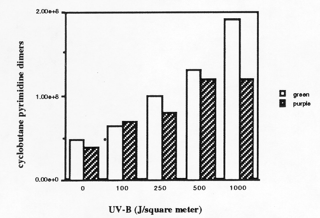
We developed a sensitive assay for two major types of UV-induced DNA damage, the cyclobutane pyrimidine dimer and the pyrimidine(6,4)pyrimidone, in DNA extracted from maize plants exposed to UV. We used chemiluminescent detection of antibodies specific to the two types of damage (Mori, Photochem. Photobiol. 54:225, 1991); we can detect as few as 107 cyclobutane pyrimidine dimers with this assay. The amount of UV-induced DNA damage in B Pl (purple) maize plants was less than the amount in isogenic b pl (green) plants (Fig. 1).
We conclude that anthocyanins protect maize DNA from UV-induced DNA damage. This suggests that anthocyanins, which are located in the epidermal vacuole, must be between the DNA and the source of UV. We used confocal laser microscopy to examine the location of the nucleus in leaf and sheath samples. In leaf epidermal cells the nucleus is in the interior third of the cell. Thus in leaves the epidermal nuclei and the mesophyll nuclei are shielded from UV by the anthocyanin in the vacuole. However, in sheath tissue the epidermal nuclei are in the outer portion of the cell; the nuclear DNA is thus not protected by anthocyanins present in the vacuole. We suggest that, in sheath, either the epidermal nuclei are more tolerant of UV damage or underlying DNA molecules (either in the mesophyll or in organelles in the epidermis that are localized below the vacuole) are protected by anthocyanin.
We are now measuring the relative levels of UV-induced DNA damage in chloroplasts, mitochondria and nuclei.
Figure
1. Number of dimers in green and purple sheath tissue irradiated with
UV-B. Isogenic B Pl and b pl maize plants were grown for
6 weeks in the greenhouse, sheath tissue was excised, irradiated with UV-B,
and DNA prepared using urea/SDS lysis buffer and phenol extraction (P.
Bedinger, P, personal communication). The DNA was slot-blotted in quadruplicate
onto Nytran, baked, blocked with 5% dried milk in TBS with 0.2% Tween-20,
then washed and reacted with the monoclonal antibody to cyclobutane pyrimidine
dimers at a 1:2000 dilution. Secondary antibody conjugated to horseradish
peroxidase was used at a 1:3000 dilution. Enhanced chemiluminescence detection
of specifically bound secondary antibody was performed according to Amersham's
protocol. The luminescence was detected on X-ray film, which was densitometer
traced. The signal from a plasmid with a known number of dimers (assayed
by T4 endonuclease V protocol; Mellon, I, PNAS USA 83:8878, 1986) was compared
with the DNA samples to determine the number of dimers in each sample.
The means of the four quadruplicate samples are presented above. A permutation
test was used to compare the four measurements from the green plants to
the four measurements from the purple plants at each UV-B dose; at P<0.1
the difference between the green and purple samples was significant in
the 250 and 1000 samples.
Return to the MNL 67 On-Line Index
Return to the Maize Newsletter Index
Return to the MaizeGDB Homepage