University of Freiburg
Tissue-specific differences of maize HMG proteins
--Klaus D. Grasser and Günter Feix
High mobility group (HMG) proteins represent a class of small (Mr 10,000-30,000) nonhistone chromosomal proteins which have been isolated from a variety of eukaryotic organisms including several plants. They have been shown to be preferentially associated with transcriptionally active chromatin and, in the case of maize and soybean, to bind to A/T-rich stretches of duplex DNA (Maier, U-G et al., Mol. Gen. Genet. 221:164-170, 1990; Jacobsen et al., Plant Cell 2:85-94, 1990). Maize HMG proteins, furthermore, exhibit a specific binding to CCAAT- and TATA- boxes of the P2 promoter of the zein gene pMS1 (Grasser, KD et al., J. Biol. Chem. 265:4185-4188, 1990).
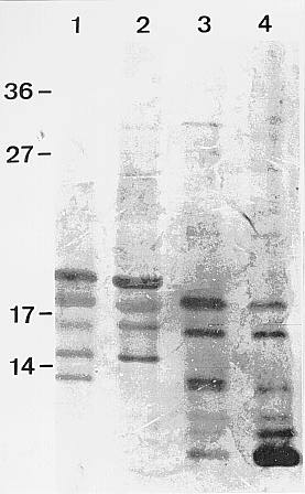
In continuation of this finding we were interested to see whether differences in the HMG protein pattern could be detected between different maize tissues and developmental stages. As shown in the figure, clear differences can be observed between HMG proteins isolated from nuclei of endosperm and leaf tissue (especially with respect to the smaller proteins visible only in leaf tissue), while only slight differences exist between the developmental stages tested. Additional evidence for differences comes from phosphorylation experiments with a casein type II protein kinase activity from endosperm nuclei (Grasser et al., Biochem. Biophys. Res. Commun. 162:456-463, 1989) which phosphorylates all the major HMG proteins from endosperm, as well as the HMG proteins of the corresponding sizes from seedling tissue, but not the smaller proteins which are unique to leaf extracts. The HMG proteins of 60 day old leaves, however, can be phosphorylated only very weakly compared to HMG proteins of other tissues. Differences were also evident in western blots with an antiserum against the small proteins from seedling tissue which does not cross-react with any HMG protein from the endosperm preparation. However, antisera against the two larger HMG proteins, both from endosperm or leaf tissue, exhibit strong cross-reactions indicating a close immunological relation. Work is currently in progress to clone the genes coding for the HMG proteins of maize. Hopefully, this will allow a more detailed analysis of the function of the HMG proteins in gene regulation.
Figure.
Silver stained protein pattern of SDS-PAGE separated HMG proteins prepared
from 7 and 14 dap endosperm tissue (lanes 1 and 2 respectively), from seedlings
(lane 3) or 60 day old leaves (lane 4). Numbers on the left indicate molecular
weight markers in kDa.
Return to the MNL 65 On-Line Index
Return to the Maize Newsletter Index
Return to the MaizeGDB Homepage