St. Mary's College
ST. PAUL, MINNESOTA
University of Minnesota
Maternal effect leads to DNA replication pattern in maize endosperm
--R. V. Kowles, G. L. Yerk, R. L. Phillips, and F. Srienc
Within two to four hours after fertilization the endosperm nucleus begins rapid synchronous divisions resulting in a syncitial tissue. Cell walls are synthesized and laid down at about four days after fertilization leading to a cellular, uninucleate tissue. Mitotic activity in the endosperm peaks at 8 to 10 dap. DNA synthesis in the endosperm peaks at 16 to 18 dap and then declines. Starch and zein synthesis in the endosperm begin at about the same time as the increase in DNA synthesis occurs.
The endosperm may be divided into three regions with regard to the DNA increase: 1) a central region in which the DNA content per nucleus is elevated, 2) a peripheral region where normal mitotic activity occurs and the nuclei remain 3C, and 3) a transitional region between the central and peripheral regions where both types of cells occur. A number of molecular tests have indicated endoreduplication as the mode of DNA increase in endosperm cells; i.e. the entire genome undergoes replication during each round. These endoreduplicating cells have a cell cycle consisting of alternating G and S phases. DNA content may reach levels as high as 384 C per nucleus in inbred A188, with an average DNA content per nucleus of 90 to 100C in the central, endoreduplicating region.
Multiparametric flow cytometry has been used to further characterize the DNA content and replication patterns of a variety of maize endosperm samples including inbreds, F1s, F2s, and F3s. Nuclear preparations made from the entire endosperm of one kernel were stained with mithramycin A. Similar preparations were made from embryo tissue. The embryo nuclei serve as 2C and 4C reference cells for the endosperm nuclei of the same line.
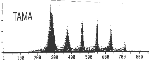
The samples were analyzed using a Cytofluorograph IIs. Size of the nuclei was measured by small-angle light scattering and DNA content was determined by the magnitude of the fluorescence signal from each nucleus. DNA replication patterns were obtained from histograms of frequency vs. fluorescence intensity processed in area mode (Fig. 1). Peaks on such a plot correspond to cells in G phase with each peak representing a doubling in DNA content from the previous one (3C, 6C, 12C, etc.). The troughs between adjacent peaks correspond to cells in S-phase. The sharpness of these peaks and the percentages of cells in the trough areas vary among inbred lines.
Analysis of pairs of inbred lines and their reciprocal hybrids revealed that the DNA replication pattern of the hybrid is the same as that of the female parent in the cross. Data from F2 kernels derived from reciprocal F1s gave DNA replication patterns among F2 kernels which were all the same regardless of the direction of the cross. This suggests the observed phenomenon is not due to either dosage effect or organellar genomes controlling the inheritance of the DNA replication patterns. If a dosage effect were present, differences among F2 kernels from an ear should be observed. In the case of organellar DNA controlling the patterns, no difference among F2 kernels from an ear would be seen but reciprocal F2 kernels derived from F1s should give different patterns.
The data described thus far suggest that the DNA replication patterns in maize endosperm nuclei could be controlled by maternal effects. Maternal effects arise due to protein(s) or transcript(s) encoded by the nuclear genome of the maternal parent that influence the development/performance of the progeny. If maternal effects are causing the observed patterns, the following outcomes would be predicted. First, the hybrid will always have the same DNA replication pattern as the maternal parent in the cross. Second, all F2 kernels will have the same pattern regardless of which direction the F1 hybrid they were derived from was made. This is because the nuclear genotype of the F1 is the same regardless of the direction of the cross. Third, provided kernels from enough different F3 ears are sampled, different patterns should be observed. This is because the F2 plants upon which the F3 seeds are growing have different genotypes.
Comparison of DNA replication patterns from F3 kernels demonstrated differences in the patterns among kernels. These findings support maternal effect as the basis of the observed DNA replication patterns.
Figure
1. Number of nuclei vs. log DNA fluorescence in arbitrary units for
self pollinated Tama Flint at 16 dap.
Return to the MNL 65 On-Line Index
Return to the Maize Newsletter Index
Return to the MaizeGDB Homepage