State University of New York
CHAMPAIGN, ILLINOIS
United AgriSeeds Inc.
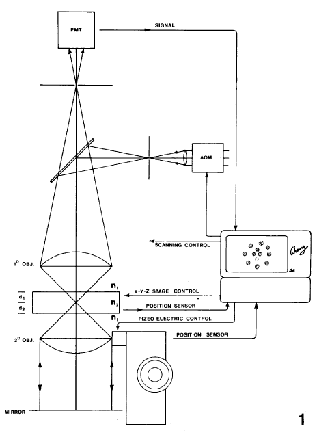
The principle of confocal microscopy is that objects in the focal plane of the objective lens are illuminated by a point source and the light reflected or emitted by the specimen is seen by a point detector (Figure 1). The key element of confocal microscopic design is a small, diffraction limited aperture positioned at the secondary focus of the objective lens in the reflected light path. The size of this aperture is such that the virtual image at the sample is of the same order of size as the focal spot. In other words, a diffraction limited illumination spot is formed on the sample. In practice, the point source and the point detector are obtained by placing apertures between a light source, detector and objective lens. Confocal imaging is achieved when the system is aligned precisely so that rays from the source aperture pass through the viewing aperture. Rays that emerge from objects out of the focal plane are not focused at the viewing aperture and are thus blocked from reaching the detector. The result is a high contrast image of a small portion of the specimen at the focal plane. To see an entire field, a means must be derived to scan either the specimen or the illumination and detector. At the present time, biological confocal microscopes can be subdivided into two categories: (1) Multi-beam scanning (e.g. Tandem scanning [A. Boyde, 1985, Proc. of RMS, 20(3):131-139] and Single-sided scanning confocal microscopes [P. C. Cheng et al., 1989, Proc. EMSA, 136-137]) and (2) Single-beam scanning (e.g. Laser scanning confocal microscope). Figure 1 shows the principle of a confocal microscope. A 50/50 beam splitter replaces the dichroic beam splitter and barrier filter when the microscope is to be operated in epi-reflective mode.
The confocal microscope has the advantage over conventional, field illumination microscopes in that it enables the contrast capabilities, not available in the conventional system, to be exploited. One of the prime advantages is the capability of optical section tomography, opening the possibility of noninvasive imaging of 3D structures with exceptional contrast and the ability to rapidly acquire and reconstruct the 3D information. Confocal microscopy can be expected to have a considerable impact on studies of chromosomal arrangement, cellular structure and tissue organization.
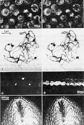
The following are some examples of the potential applications of confocal light microscopy in maize research. For practical reasons and simplicity in demonstrating the applications, a commercially available (BioRad MRC-500) laser scanning confocal microscope is used in our discussion. One of the potential applications of confocal microscopy is the study of tissue organization. Figure 2 shows a 3D image pair of the nuclei of a developing tapetum layer in a maize anther. The image was obtained from a whole mount specimen by focusing through three layers of cells (epidermis, endothecium and middle layer). Figure 3 shows a 3D image pair of chromosomes at pachytene stage of meiosis. Both Figures 2 and 3 were obtained from intact anthers which were fixed in 1:3 acetic acid-EtOH fixative, stained with Feulgen reaction, dehydrated in EtOH and cleared in methyl salicylate (to improve the optical properties of the specimen [Cheng and Summers, 1990, In: The Handbook of Biological Confocal Microscopy, ed. J. Pawley, Plenum Press]). Figure 4 shows a confocal fluorescent image of the nuclei of silica cells of maize leaf (Feulgen stained). All three images were obtained in epi-fluorescent mode by using the 514nm line of Ar ion laser as the excitation wavelength. In order to improve the fluorescent signal, four Al-coated folding mirrors in the scanning system were replaced by high reflective dielectric mirrors.
One of the major difficulties we have encountered in confocal fluorescent imaging is photo-bleaching of the fluorochromes. This is particularly problematic when numerous high resolution optically sectioned images are required. In order to further improve the signal strength and reduce photo-bleaching, we are currently working on two major hardware implementations: (1) design and construct an auto-focus, folded optics confocal trans-illumination system (TIS, optics placed below the specimen in Figure 1). Instead of operating in trans-illumination mode, the TIS can be used to increase the excitation intensity and improve the strength of the fluorescent signal (by increasing detector solid angle); and (2) an acousto-optic modulator (AOM) will be installed in the illuminating beam to minimize photo bleaching resulting from the retracing cycle of the scanning laser beam. The AOM turns off the laser beam during the retracing cycle of the two scanning mirrors, eliminating all the non-image producing exposure to the laser beam.
In addition to epi-fluorescent mode,
confocal microscopy operated in reflective (back scatter) mode can also
be very useful in maize research. For instance, Figure 5 illustrates a
back scattered confocal image of the silica deposition within dumb-bell
shaped silica cells. The reflective image was obtained simultaneously with
the fluorescent image shown in Figure 4. Furthermore, reflective confocal
microscopy can be used to study the surface of a specimen (both fresh or
fixed and dehydrated tissue). A typical example of a reflective confocal
image which shows the surface features of a critical point-dried coleoptile
is illustrated in Figure 6.
Return to the MNL 64 On-Line Index
Return to the Maize Newsletter Index
Return to the MaizeGDB Homepage