ß-glucosidase
zymograms in denaturing (SDS) gels
--Asim Esen and Gunay Gungor
We performed a systematic search for concentrations
of protein denaturing agents that inactivate maize ß-glucosidase
activity. In the course of these studies, it was discovered that the enzyme
retained about 50% of its activity after treatment with or in the presence
of the anionic detergent sodium dodecyl sulfate (SDS), a potent denaturant,
when used at or around 1% concentrations. When coleoptile extracts or purified
enzyme preparations were incubated in the presence of SDS concentrations
varying from 0 to 1.6%, electrophoresed through 6-8% polyacrylamide gels
and stained for activity, zymogram patterns similar to those of untreated
samples were obtained (Fig. 1a). However, the intensity of enzyme bands
decreased as SDS concentration increased to 0.8% or higher. In view of
these results, controls and SDS-treated samples were subjected to standard
SDS-PAGE through 10-12% gels using the method of Laemmli and the resulting
gels were stained for enzyme activity. Surprisingly, a zone of activity
including 1 to 6 bands, depending on sample age and buffer pH and composition,
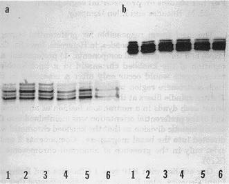
in the 60 kD region of the gel was obtained (Fig. 1b). Figure 1 shows representative
zymograms obtained when the control and SDS-treated samples were electrophoresed
through a 6% native (Fig. 1a) and a 10% SDS gel (Fig. 1b). Multiple ß-glucosidase
bands are visible in both zymograms. In this case, the source of the heterogeneity
is thought to be artifactual due to the activity of an endogenous SH-proteinase
because it occurred independently of SDS treatment and the samples were
handled under conditions (e.g., extraction and storage in a pH 5 buffer
containing 2-mercaptoethanol) promoting the activity of such proteinase
prior to SDS treatment and electrophoresis. In subsequent experiments (results
not shown), a single band was obtained when the sample was not exposed
to the reducing agent. Furthermore, it was shown that the zymograms of
the maize inbreds classified as null at the Glu locus contained
ß-glucosidase bands after their coleoptile extracts were treated
with SDS. These results suggest that the 60 kD polypeptide, the ß-glucosidase
monomer, shows full enzymatic activity and, thus, the in vivo form of the
functional enzyme is a monomer, not dimer as assumed on the basis of zymogram
patterns of hybrid genotypes. In addition, the results clearly show that
the maize inbreds that are classified as null for Glu encoded ß-glucosidase
activity are not null. The enzyme of these so-called null genotypes appears
to occur as large aggregates that fail to enter the gel and cannot be detected
by standard zymogram techniques.
Figure
1. ß-glucosidase zymograms of a maize coleoptile extract after
treatment with SDS prior to electrophoresis.
a, 6% nondenaturing
(native) gel. b, 10% denaturing (SDS) gel. The enzyme was extracted
with 25 mM NaAc buffer, pH 5.0, containing 35 mM 2-mercaptoethanol. Lane
1, control (no SDS added). Lanes 2-6, after adding SDS to a final concentration
of 0.1 to 1.6%. 2, 0.1%; 3, 0.2%; 4, 0.4%; 5,0.8%; 6, 1.6%.
Please Note: Notes submitted to the Maize Genetics Cooperation
Newsletter may be cited only with consent of the authors
Return to the MNL 64 On-Line Index
Return to the Maize Newsletter Index
Return to the MaizeGDB Homepage