--Ted Klein, Brad Roth and Michael Fromm
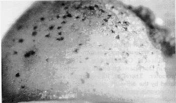
We have employed microprojectiles to deliver the A1 and Bz1 genes to cells within intact aleurone tissue. Aleurones from plants carrying C1 and R and either a1 or bz1 were excised 15 days after pollination by peeling back the pericarp and removing the aleurone and underlying endosperm tissue. The tissue was placed on agar-solidified medium and bombarded with microprojectiles (Klein et al., Bio/Technology 6:559, 1988) coated with the genomic clones of the A1 (C. O'Reilly et al., EMBO J. 4:877, 1985) or Bz1 (N. Fedoroff et al., Proc. Natl. Acad. Sci. USA 81:3825, 1984) genes. After incubating the tissue for 1 day in the light at 26 C, a portion of the cells within the aleurone developed purple pigmentation (Fig. 1). Individual isolated purple cells could be visualized but more generally 3 to 5 adjacent cells developed pigmentation with a central most cell being more highly pigmented than surrounding cells. These pigmented spots were restricted to the aleurone layer. Up to 100 of these spots developed on a single a1,C1,R or bz1,C1,R aleurone following bombardment with either the A1 or Bz1 genomic clone, respectively. Such spots did not develop when aleurone tissue was bombarded with microprojectiles coated with pUC18. This indicates that the introduced A1 or Bz1 genomic clones complemented the a1 and bz1 mutations and that the isolated clones are functional when reintroduced into aleurone cells.
Figure 1. Expression of the Bz1 genomic clone following its delivery into maize aleurone tissue from bz1,C,R plants. Anthocyanin pigment appears as dark spots on the black-and-white photograph.
To analyze the expression of these genes in different genetic backgrounds, chimeric genes were constructed by fusing the firefly luciferase coding region to the 5' and 3' regions from the A1 or Bz1 genomic clones to form pA1L and pBz1L, respectively. These constructs were introduced into aleurones of the following genotypes: C1,R; c1,R; C1,r; c1,r; C-I,R. Levels of luciferase activity in permissive backgrounds (C1,R) were about 20 to 100 fold greater than those detected in tissue carrying either or both of the recessive alleles (c1,r) of these genes (Table 1). Low levels of luciferase were also observed following delivery of pA1L or pBz1L to C-I,R aleurones. As a positive control, aleurones from the various genotypes were bombarded with pAI1LN (J. Callis et al., Genes Develop. 1:1183, 1987). The luciferase gene in this plasmid is under the control of the maize Adh1 promoter. Therefore, its expression should not be influenced by the different genetic backgrounds that regulate anthocyanin production. Levels of luciferase activity were consistently high in aleurone tissue from all of the genotypes tested following delivery of pAI1LN-DNA.
Table 1. Luciferase expression in aleurone tissue from various genotypes
following bombardment with the chimeric A1 or Bz1 genes.
| GENOTYPE | PLASMID | LUCIFERASE ACTIVITY + S. E. (light units x 103)* | |
| a1, C1, R | pA1L | 20.4 + | 3.8 |
| pBz1L | 26.1 + | 8.1 | |
| pAI1LN | 124.4 + | 21.9 | |
| bz1, C1, R | pA1L | 22.6 + | 0.3 |
| pBz1L | 33.3 + | 11.2 | |
| pAI1LN | 319.0 + | 20.4 | |
| c1, r | pA1L | 0.5 + | 0.2 |
| pBz1L | 0.2 + | 0.01 | |
| pAI1LN | 141.1 + | 56.4 | |
| c1, R | pA1L | 1.2 + | 0.2 |
| pBz1L | 0.6 + | 0.1 | |
| pAI1LN | 193.5 + | 23.4 | |
| C1, r | pA1L | 0.3 + | 0.1 |
| pBz1L | 0.5 + | 0.3 | |
| pAI1LN | 299.2 + | 85.6 | |
| C-I, R | pA1L | 1.2 + | 0.1 |
| pBz1L | 0.3 + | 0.0 | |
| pAI1LN | 446.2 + | 168.4 | |
*Luciferase activity was determined according to Callis et al. (Genes Develop. 1:1183, 1987).
These results show that expression of DNA delivered to intact tissues
by microprojectiles properly reflects the regulation of the native genes.
The transfer of genes directly to intact tissues provides a rapid means
for studying the genetic regulation of gene expression at the molecular
level.
Return to the MNL 63 On-Line Index
Return to the Maize Newsletter Index
Return to the MaizeGDB Homepage