There is no vascular connection between the maternal plant and developing caryopses of corn. All nutrients entering the seeds are believed to pass through, by symplastic or apoplastic methods, a specialized group of cells called transfer or basal endosperm cells. Even though these cells are of primary importance in the process of grain filling, surprisingly little is known about their biochemical and physiological processes. During ongoing studies in our laboratories concerning the physiology of grain filling, it became apparent that the few previously published descriptions of transfer cells in corn were not adequately describing what we were seeing in our own anatomical investigations.
Sections were made, for examination under light and electron microscopes, of various stages of caryopsis development of Tx5855, starting at four days post pollination and proceeding until development of a black layer. Samples of grains at 23 days post-pollination were chosen to illustrate the structure of mature, functioning transfer cells. The samples were examined in two planes of section, one along the long axis of the cells and referred to as longitudinal, the other perpendicular to the long axis and referred to as cross section.
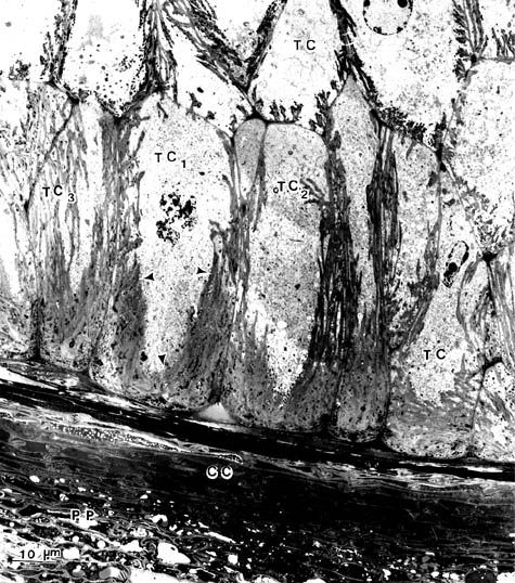
These two planes of section showed that the typical transfer cell had an extensive network of cell wall ingrowths, all of which were bounded by plasma membrane. In longitudinal sections (Fig. 1) the transfer cells had the greatest cell wall proliferation in the basal portions of the cells and progressively less toward the apices. The central regions of the cells were devoid of cell wall material and were filled with cytoplasm containing normal organelles including numerous vesicles and occasional crystals. The overall appearance of these cells was goblet-shaped.
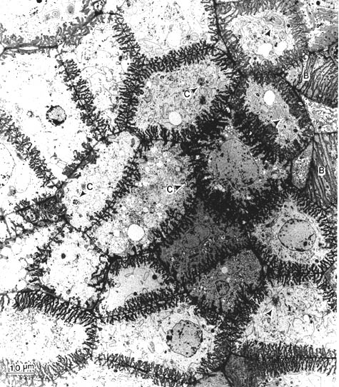
In cross section the extension of the ingrowths from the primary cell wall could be easily observed (Fig. 2). One of the most striking features found in this plane was that wall ingrowths of contiguous cells often arose from a common site on the primary wall. In the areas between the ingrowths, numerous plasmodesmata were present.
The most basal transfer cells had the greatest amount of cell wall proliferation. Evidence of wall extensions could be found in cells approximately 6 layers deep in the endosperm. Each successively deeper cell layer had less proliferation until there was a complete transition to typical endosperm cells. There was also a transition area at the lateral edges of the transfer cell zone where it meets the aleurone layer.
An hypothesis currently under our consideration is that the production of the transfer cell zone is initiated by a diffusible promoter, possibly a hormone. The primary effect of this substance is in the most basal endosperm cells. These are the first non-maternal cells that assimilates come in contact with. As the promoter substance diffuses inward it may have progressively less effect, accounting for the eventual transition from transfer to endosperm cells. The production of the aleurone layer is presumably also under hormonal control. The competition between the initiating substances for the aleurone and transfer cell zones might account for the transition zone where these tissues meet.
Figure 1. This transmission electron micrograph shows transfer cells in a longitudinal section, crushed nucellar cells, and pedicel parenchyma from a corn caryopsis 23 days post pollination. Note the basal transfer cells have extensive cell wall proliferation at the base and along the sides (unlabeled arrows) and that the centers are filled with cytoplasm. This gives these cells a goblet-like appearance (TC1, TC2). Other transfer cells are present in this layer that do not have the goblet appearance because the section plane is off median (TC3) TC: Transfer cell; CC: crushed cells; PP: pedicel parenchyma
Figure 2. This transmission electron micrograph shows transfer cells in cross section. Note that most of the contiguous cells have wall extensions originating at common loci. This plane of section is probably through either the upper portion of basal transfer cells or through second level transfer cells. The basal portions of two second level cells are present (B). Some of the cells contain an unusual aggregation of endoplasmic reticulum (unlabeled arrows) and plastids with crystalline inclusions (arrows labeled C).
Ronald W Davis, B. Greg Cobb and J.D. Smith
Return to the MNL 62 On-Line Index
Return to the Maize Newsletter Index
Return to the MaizeGDB Homepage