The embryo-lethal mutant dek1 shows pleiotropism, since, in addition to the block of embryogenesis at the proembryo stage, the endosperm is white, being devoid of carotenoids and anthocyanins, while the aleurone layer is absent. With the aim of better characterizing this mutant, two nuclear parameters, DNA replication and DNA content, were analyzed and the data obtained submitted for publication.
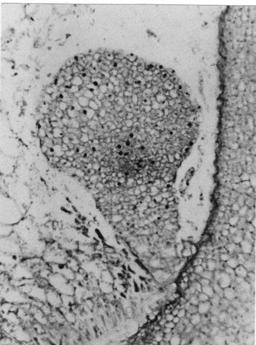
The capacity of mutant embryonic cells to actively synthesize DNA, in spite of the arrest in differentiation, was tested by determining the 3H-thymidine incorporation in 14 DAP kernels. Normal and mutant sib kernels were cultured for 48 h in sterile MS liquid medium supplemented with 30µCi/Ml 3H-thymidine (specific activity 51µCi/mmol) or alternatively injected with 5 µCi 3H-thymidine and then cultured in MS medium for 48 h. Both procedures adopted gave positive results. Autoradiographic silver grains were preferentially located over nuclei of the central differentiated area in the mutant embryo, even though incorporation is also observed in nuclei located in the peripheral region (Fig. 1). Cell division capacity therefore is not affected by the mutation, as is also shown by the possibility of inducing callus from mutant embryos.
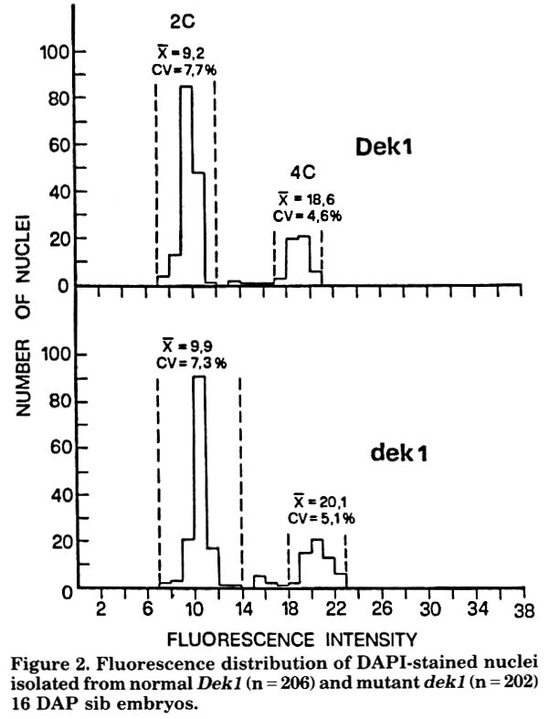
On the other hand, the increase in nucleolus and nucleus size observed both in embryo and in endosperm mutant cells (MNL 61:73, 1987) raised the question of whether larger nuclei might be the result of a higher DNA content and larger nucleoli of a consequently larger number of transcribed rRNA genes. DNA quantification by means of cytofluorimetry was therefore performed on nuclei isolated from normal and mutant embryonic cells at 16 DAP The overlapping of the two fluorescence distributions of DAPI-stained nuclei (Fig. 2) leads one to exclude an increase in DNA content in mutant cells. Further autoradiographic experiments with 3H-thymidine and 3H-uridine incorporation are in progress.
Figure 1. Autoradiograph of a longitudinal section of a 14 DAP mutant embryo after incubation in MS medium supplemented with 3H-thymidine (30µCi/ml) for 48 h. Labelled nuclei appear as black dots over cells. X 135
Figure 2. Fluorescence distribution of DAPI-stained nuclei isolated from normal Dek1 (n = 206) and mutant dek1 (n = 202) 16 DAP sib embryos.
Silvana Faccio Dolfini, Francesca Sparvoli and Helene Libert
Return to the MNL 62 On-Line Index
Return to the Maize Newsletter Index
Return to the MaizeGDB Homepage