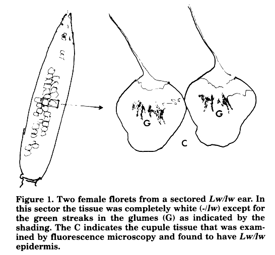
We are using X-ray induced marked somatic clones to study floral development in plants carrying the Miniplant (Mpl) mutation. Mpl is a mild dominant dwarfing mutation, possibly allelic to D8, causing the formation of anthers in the florets of the ear. The clones are marked with the absolutely white phenotype conferred by the recessive lemon-white (lw) allele approximately 60 map units out on the long arm of chromosome 1. Mpl is also on this arm and is approximately 4 map units distal to lw. Plants heterozygous for lw (Mpl Lw/+ lw) are irradiated (X-ray, 1000R) shortly after germination. Tissue derived from cells that have lost a region of the chromosome arm carrying the functional (Lw) allele are seen as white sectors in the mature plant. Each sector represents a patch of tissue developed clonally from a single cell. We have observed a number of sectors in ear husks. These husks were removed in order to permit greening of the florets, cupules and stem tissue beneath. In the normal ear exposure to light causes greening of the stem, cupules and glumes, just as it does in the homologous organs of the tassel. We identified several lw sectors which extended into the floral tissues resulting in completely white glumes, cupules and stem. These have enabled us to determine that the Mpl character is cell autonomous (manuscript in preparation). We also observed sectors which were almost completely white in appearance except for the presence of some narrow green streaks beginning at the glume tips and radiating outwards and downwards (Figure 1). Analysis of the white cupule tissue adjacent to these florets by fluorescence microscopy (illuminated at 395-440nm, fluorescence observed at 470nm) revealed the presence of epidermal guard cells containing chlorophyll indicating that this region consisted of a -/lw mesophyll covered by a Lw/lw epidermis.
These observations suggest the occurrence of programmed periclinal cell divisions of the epidermis during glume development. These divisions into the plane of the glume result in Lw/lw internal cells derived from the epidermis. The divisions are programmed because the green streaks are found in glumes of 15-20 adjacent florets in the same sector. They are programmed loosely because the streaking pattern in each floret is similar but not identical. It is interesting to note that the tip of the leaf, a homologous organ to the glume, is almost entirely derived from periclinal divisions of cells in the epidermal layer. This similarity between the glume and the leaf tip was pointed out to us by Scott Poethig. Maybe the differences in spatial organization of these epidermal divisions represent'glume' and 'leaf variants of homologue specific epidermal cell division programmes.
Figure 1. Two female florets from a sectored Lw/lw ear. In this sector the tissue was completely white (-/lw) except for the green streaks in the glumes (G) as indicated by the shading. The C indicates the cupule tissue that was examined by fluorescence microscopy and found to have Lw/lw epidermis.
Nicholas Harberd, Sarah Hake and Michael Freeling
Return to the MNL 61 On-Line Index
Return to the Maize Newsletter Index
Return to the MaizeGDB Homepage