The embryo-lethal recessive 561 mutant, isolated in our lab following EMS mutagenesis and shown to be allelic to dek1 (W F Sheridan, M. T. Chang and M. G. Neuffer, MNL 58:98-99, 1984), is characterized by defects both in embryo and in endosperm development, absence of a correctly differentiated aleurone layer and suppression of both carotenoids and anthocyanins. Here a description is given of the cytology of endosperm and embryo tissues of the mutant dek1.
Embryos and endosperms of four mutant and four normal sib kernels collected 16 days after pollination (DAP), when the mutant is first recognizable, were separately fixed. The procedure followed to obtain cytological preparations was that described by B.-Y. Lin (Stain Technology 52:197-201, 1977) with slight modifications. Dissociation into cells was achieved mechanically by needles and preparations were obtained by pipetting a few drops of the cell suspension onto clean slides warmed on a hot plate. Silver staining (50%AgNO3 at 60 C) was applied in order to visualize nucleolar structures.
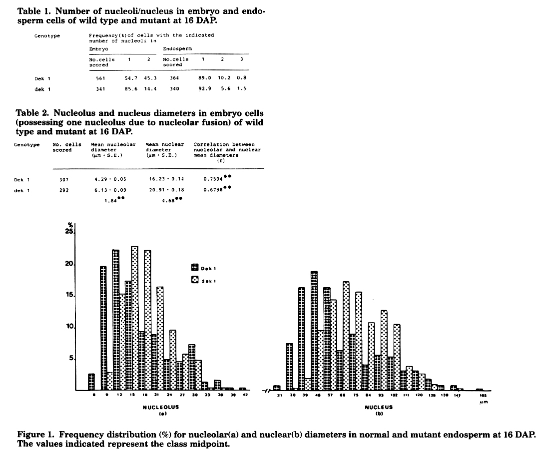
The number of nucleoli per nucleus in normal and mutant kernels is shown in Table 1. The results are strikingly different in the two tissues. In the embryo, in fact, the frequency of cells with two nucleoli in the mutant is significantly lower than that found in non-mutant embryos, the majority of cells being with one nucleolus. In the endosperm, on the other hand, the distribution of nucleoli among the three frequency classes is apparently equal in both mutant and normal. Keeping in mind that endosperm cells at the time of observation (16 DAP) are no longer dividing, these results suggest a total or partial impairment of mutant embryo tissues to go through active divisions.
Nucleolar and nuclear diameters for both embryo and endosperm were measured by an eye piece micrometer in cells displaying one nucleolus. The data pertaining to embryo measurements (Table 2) indicate a significant increase in size of the mutant vs. normal embryos. Similar conclusions are obtained in endosperm cells (Fig. 1), where a shift of the mutant frequency distribution towards higher values, if compared to the non-mutant values, is observed.
These preliminary data are consistent with the hypothesis of a difference in kinetics of the two cell populations and further studies will be aimed at the elucidation of these aspects.
Silvana Faccio Dolfini
Return to the MNL 61 On-Line Index
Return to the Maize Newsletter Index
Return to the MaizeGDB Homepage