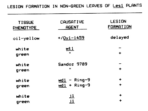
Lesion expression in various types of non-green leaves of +/Les1 plants has been examined (see table). Lesion expression in leaves with a low chlorophyll content due to Oy1-1459 is delayed over normal green sibs by approximately 2-3 weeks. When lesions do form, they are larger and generally more banded in appearance. Since it is unclear whether photosynthesis is required for lesion formation (see article by C. Echt), the delayed lesion formation may be due to a slower accumulation of some metabolite which is required to trigger lesion formation or is involved in the production of visible necrosis.
Lesion formation was also examined in various white sectors in leaves. Lesions were never seen to initiate or enlarge into the white tissue of wt1 seedlings or white leaves following treatment of seedlings with the herbicide Sandoz 9789. Lesions forming in the green tissue near the white border never enlarged into the white tissue. These findings seem to indicate that lesion formation and enlargement requires photosynthesizing tissue; however, lesions were found to form in the white stripes of j1 (japonica) and wd1 + Ring-9 (Wd1 C-1) plants. There was some indication that lesion formation in the white and perhaps layered sectors of the wdl plants was more pronounced than in adjacent green tissue. Lesion formation in non-sectoring green sibs of wd1 plants was also lower, perhaps indicating that sectored and/or layered leaf tissue is more primed to initiate lesions in the presence of Les1. Pure white seedlings resulting from the complete loss of the ring chromosome never formed lesions. These seedlings survived for only 2-3 weeks and lesion formation may require a longer period since the Oy1-1459 plants were delayed in their expression. It is unclear why lesions formed in one type of white tissue and not the other. Perhaps longitudinal sectors such as those formed by j1 and wd1 are cross-fed by neighboring green tissue differently than lateral divisions of white and green tissue. This feeding may provide a necessary metabolite for lesion formation. It is also possible that the layering, which is absent in wt1 and Sandoz tissue, is responsible for lesion formation.
Dave Hoisington
Return to the MNL 60 On-Line Index
Return to the Maize Newsletter Index
Return to the MaizeGDB Homepage