Silver-staining methods, developed for localizing nucleolar organizer regions (NORs) both in animal and plant chromosomes, reveal gene activity at the ribosomal DNA sites. The procedure described by M. Hizume et al. (Stain Technology 55:87-90, 1980) was applied to maize root tip cells with some modifications. Pretreatment, fixation and washing are performed as described in the cited paper. Differences are as follows:
- Each step is carried out on a single root tip placed in a small container
- After washing off the enzyme solution by several changes of distilled water, a few drops of 60% acetic acid are added for 30 minutes
- Dissociation into cells is achieved mechanically by needles
- Chromosome permanent preparations are obtained by pipetting a few drops of the cell suspension onto clean slides warmed on a hot plate (40-45 C)
- For Ag-staining, slides treated with 50% AgNO3 and covered with a coverglass are kept in a moisture chamber for one hour at 60 C and illuminated by a 60W bulb.
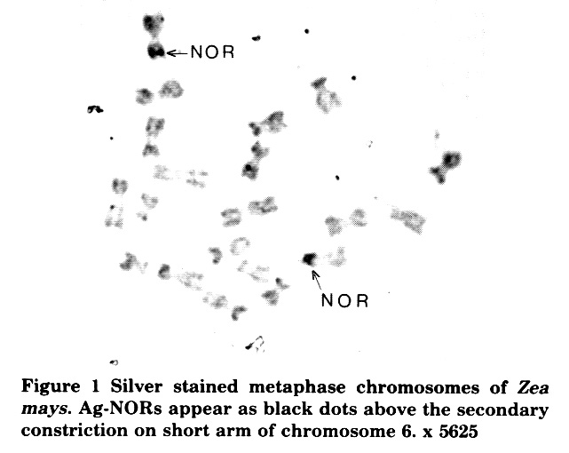
Figure 1 shows root tip metaphase chromosomes prepared and stained as described: NORs are clearly visible on fhe short arm of chromosome 6. The possibility of visualizing ribosomal gene activity offers many opportunities in cytogenetics and genetics.
Figure 1. Silver stained metaphase chromosomes of Zea mays. Ag-NORs appear as black dots above the secondary constriction on short arm of chromosome 6. x 5625
Silvana Faccio Dolfini
Return to the MNL 60 On-Line Index
Return to the Maize Newsletter Index
Return to the MaizeGDB Homepage