Successful application of tissue culture technology provides powerful tools to supplement conventional breeding methods in crop improvement. However, such success depends not only upon the ability to regenerate plants in vitro but also upon the ability to grow our plants in vitro as model systems. Cells cultured in vitro as calli are able to regenerate plants in two ways: by a sequential differentiation of shoots and roots which by association with each other form plants, or by somatic (nonzygotic) embryogenesis. In the first case, organogenesis, shoot and root meristems are multicellular in origin. On the other hand, somatic embryogenesis arises apparently from single cells. The establishment of a more or less dedifferentiated tissue culture under defined culture conditions gives rise to a subsequent regeneration of plants which frequently show phenotypic alterations. Plants of multicellular origin cannot always be expected to be genetically uniform. However, plants obtained through somatic embryogenesis are suitable for mutant research, genetic analysis, maintenance of genetic stocks, and breeding (Vasil, I. K., Cell & Tiss. Cult. Tech. for Cereal Crop Improv., 131-144, 1983).
In maize, it is now possible to obtain plants either through organogenesis or from somatic embryogenesis. In our work, the primary genotypes used included several floury-a inbred lines, red flint inbred lines and hybrids between them. The experiments were carried out in the IFSC (Buenos Aires, Argentina) during the spring of 1983. Ears were removed from greenhouse-grown plants at 12 to 18 days after pollination. The kernels were taken from the middle part of the ears and then were sterilized for 20 min in 2.5% sodium hypochlorite solution and rinsed twice (5 min each) with sterile water before culture. Immature embryos were isolated from the kernels and were then placed on the solid culture medium with the plumule-radicle axis in contact with the medium and the scutellar side exposed. The primary cultures were incubated at 28 C in the dark.
Calli were initiated on three different media:
Medium A: Inorganic components of Murashige-Skoog, organic components of Straus, plus 20 g sucrose/liter, 8 g agar/liter and 1 mg 2,4-D/liter.
Medium B: Medium A plus 120 g sucrose/liter, 400 mg proline/liter, 8 g agar/liter and 1 mg 2,4-D/liter.
Medium C: Yu-pei medium plus 120 g sucrose/liter, 400 mg proline/liter, 500 mg casein hydrolysate/liter, 8 g agar/liter and 1 mg 2,4-D/liter.
The frequency of positive culture response from immature embryos of all genotypes ranged from 60 to 100%. Calli were white to pale yellow in coloration. However, the texture of the calli obtained in the three media differed markedly. The variation of the texture of the calli among media was markedly higher than the variation among genotypes in each medium.
Medium A is the common medium developed by Green and Phillips and used by several authors to culture maize in vitro. Calli obtained in such medium were friable and nodular. After 30 days, these primary cultures developed 1 to 1.5 cm good callus masses. Medium B is a variation of medium A, with a high osmotic concentration and proline level. Medium C is a medium developed by Chinese scientists to obtain androgenesis in maize. The textures of the calli obtained in medium B and C were highly similar to each other. Culture initiation began with enlargement of the scutellar surface, which resulted in a very prominent domeshaped, compact and smooth scutellum, with continued growth. However, these scutellum calli obtained in medium B and C proliferated more slowly than calli obtained in medium A.
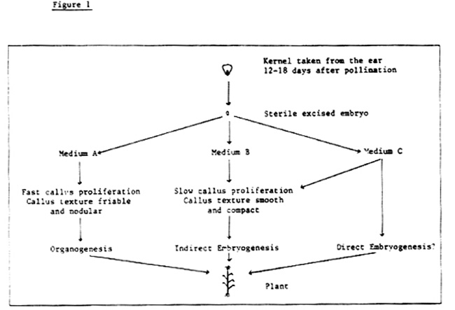
The morphogenetic events observed in the three media were different (Rapela, this MNL). These facts let us suppose that it is possible in our material to induce both organogenesis and embryogenesis with an adequate change of the medium constituents. Also we will introduce evidence of direct embryogenesis in some cultures obtained in medium C. We suggest for the maize material cultured under our conditions a tissue culture model experimental system as shown in Figure 1.
Miguel Angel Rapela
Return to the MNL 58 On-Line Index
Return to the Maize Newsletter Index
Return to the MaizeGDB Homepage