In the preceding paper we demonstrated that maize kernel weight is influenced by incompletely dominant genetic factors present on chromosomes 2 and 7. We also determined the effects of trisomy on embryo volume by comparing the embryo volumes of sibling trisomic and disomic kernels. In this way, most of the maize genome was screened for incompletely dominant genetic loci that influence embryo volume.
The kernels employed in this study were the same kernels analyzed in the previous investigation of kernel weight. In addition, the same designation of kernels into families was maintained throughout these experiments.
After the weight of the kernels was determined, they were scanned with a nuclear magnetic resonance (NMR) spectrometer. The results of the NMR spectroscopy analyses will be presented elsewhere. Following NMR spectroscopy, the kernels were placed at 100% humidity for 24 hours. This hydration was necessary to soften the kernels for slicing, which was part of the embryo measurement procedure. The measurements were made immediately after the kernels were removed from the humid environment.
The method used to estimate the embryo volumes was that developed by Plewa (Plewa and Weber, 1973, Can. J. Genet. Cytol. 15:313). A dissecting microscope equipped with an ocular micrometer was used at 10X magnification to determine the embryo dimensions. First, the length and width of the surface of the embryo of each uncut kernel was measured. Each kernel was then cut in half parallel and adjacent to the embryonic axis. The portion of the kernel containing the embryonic axis was turned on its side and the depth of the embryo was measured. Each measurement was made by determining the greatest linear distance within the boundaries of the scutellum.
The product of length times width times depth was calculated for each kernel; this product is an estimate of embryo volume. All determinations of embryo volumes were made prior to the identification of trisomic kernels and thus, experimental bias was absent from this study.
Kernel halves containing the embryonic axis were germinated and root-tips were collected from the resultant plants. These root-tips were used in somatic chromosome counts as described in the preceding report in this Newsletter. Unfortunately, the entire family segregating for trisomy of chromosome 9 was lost during germination due to rodent predation; thus no data are available from trisomic-9 kernels.
Plants cytologically identified as trisomic were transplanted to the field and allowed to grow to maturity. Microsporocyte samples were removed from trisomic plants in each family and analyzed cytologically to confirm the identity of the trisomic chromosome.
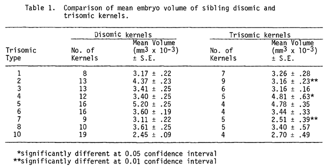
In each family of sibling trisomic and disomic kernels, the embryo volume estimates (hereafter designated as embryo volume) of trisomic and sibling disomic kernels were compared. These comparisons were made using Student's t-test. The results of these comparisons are presented in Table 1.
The effects of trisomy of different chromosomes on embryo volumes were highly specific. Disomic kernels had larger embryo volumes than their trisomic siblings in six of the nine trisomic types studied. Thus, the volumes of embryos in the trisomic kernels are generally smaller than in their disomic sibling kernels.
When the mean embryo volumes for trisomic kernels were compared with those for their sibling disomic kernels, significant differences were found for three trisomic types analyzed. The embryo volume of trisomic 2 kernels was significantly smaller (p < 0.01) than their disomic siblings. Trisomic 2 kernels had a mean embryo volume of 3.16 mm3 compared to 4.37 mm3 x 10-3 for their disomic siblings. Trisomic 7 kernels also had embryo volumes that were significantly smaller (p < 0.01) than their disomic siblings; the respective mean embryo volumes were 2.51 mm3 x 10-3 and 3.11 mm3 x 10-3. In contrast, kernels trisomic for chromosome 4 had a mean embryo volume of 4.81 mm3 x 10-3, while sibling disomic kernels' mean embryo volume was 3.40 mm3 x 10-3. This difference was also significant (p < 0.05).
Because kernels that were trisomic for chromosomes 2, 4, or 7 had embryo volumes that were significantly different from those for their respective disomic siblings, incompletely dominant genetic factors that influence embryo volume reside on these chromosomes.
It is interesting to note that an earlier investigation (Plewa and Weber, 1973, Can. J. Genet. Cytol. 15:313) employing maize kernels monosomic for chromosomes 2, 7, 8, or 10 showed that monosomic 10 embryos were significantly smaller than diploid control embryos. In the current study, trisomic 10 embryos were only slightly, but not significantly larger than disomic kernels. Plewa and Weber's study found that the volumes of monosomic 2 and monosomic 7 embryos were not significantly different from disomic control embryos. Trisomic 2 and trisomic 7 embryos were found to be significantly smaller than disomic sibling embryos in the present study. The reasons for the differences in these two studies are not known, but they might be attributed to strain differences in the maize stocks employed. In addition, monosomic embryos are accompanied by euploid (triploid) endosperm whereas trisomic embryos are surrounded by aneuploid (pentasomic) endosperm. The trisomic chromosome in the embryo is present in five copies in the endosperm. Perhaps the aneuploid endosperm of trisomic kernels exerts some influence over embryo volume.
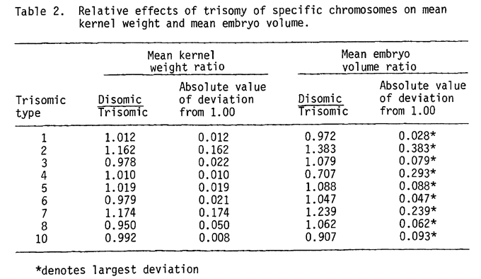
A comparison can be made of the relative effects of trisomy of specific chromosomes on kernel weight (discussed in the preceding paper) and on embryo volume. To determine the effects of trisomy of a specific chromosome on kernel weight, the ratio of mean disomic kernel weight to mean sibling trisomic kernel weight was calculated for each family. Similarly, the ratio of mean disomic embryo volume to mean sibling trisomic embryo volume was calculated for each family. These ratios are presented in Table 2.
A comparison of the absolute values of the deviation from 1.00 of the kernel weight ratios and the embryo volume ratios for each trisomic type can be used as an index of the magnitude of the effect of trisomy on these kernel parameters. The deviation of the embryo volume ratio is greater than the deviation of the kernel weight ratio for all but one of the trisomic types analyzed. Hence, the effect of trisomy was greater on mean embryo volume than on mean kernel weight for all nine chromosomes tested.
Walter D. Fox and David F. Weber
Return to the MNL 51 On-Line Index
Return to the Maize Newsletter Index
Return to the MaizeGDB Homepage