Maize Genetics Cooperation Newsletter vol 84 2010
Please Note: Notes submitted to the Maize Genetics
Cooperation Newsletter may be cited only with consent of authors.
DNIEPROPETROVSK, UKRAINE
Institute of Grain Farming, UAAN,
Ukrainian State University of Chemistry and Technology
The
development of isolated maize caryopses in vitro
Satarova, TN; Lyapustina, OV
Technique of in vitro development of isolated plant generative structures from early stages to maturity is little processing, but perspective experimental system for modeling the development and maturation of embryo sac, pollen grain, embryo, endosperm, seeds, for recovering key stages and limiting factors in formation of reproductive organs, which for many agricultural crops generate yield and seed productivity. The development of different isolated generative structures grown in vitro up to maturity is an important step in genetic transformation procedures with egg cells, sperms, zygotes, young embryos, in manipulations with single cells, in transferring organelles to gametes and zygotes.
In vitro culture of maize kernels attached to cob tissue were used in investigations of carbohydrate and protein metabolism and phytohormonal regulation of kernel development (Misra, S, Oaks, A, Plant Physiol. 77: 520-523, 1985; Cobb, BG et al., Annals of Botany 62: 265-270, 1988; Singletary, GW, Below, FE, Plant Physiol. 89: 341-346, 1989; Hole, DJ et al., Plant Physiol. 91: 105-107, 1989). Culture of maize caryopses isolated from the cob was not established yet. The objectives in the present study were to investigate effects of genotype and culture duration on the in vitro development of isolated maize caryopses and embryos inside them.
Caryopses from field donor plants of maize inbred DK366 and hybrid A22xDK307 were isolated from ear cob at the 5th-7th day after pollination. They were sterilized in 70o alcohol for 1-2 seconds, then washed three times in sterile water and explanted on nutrient medium NBM (M�l, R et al., Planta 189: 213-217, 1993) with 90 g/l sucrose and 1 mg/l 6-benzylaminopurine. Caryopses were incubated in Petri dishes at 26oC in darkness. The results of cultivation were estimated at the 20th and the 50th days in culture. Starch in different parts of kernels was determined in the reaction with Lugol�s solution. Well-developed embryos were isolated out of cultivated caryopses and transplanted for germination on modified Murashige & Skoog medium containing twice-reduced concentration of macro- and microsalts.
By the moment of explantation caryopses of investigated genotypes were of 5 mm long, white coloured (Figure 1, A), without starch in covers. Embryo sacs inside caryopses were of about 1,2-1,7 mm long and each contained an embryo of about 0,1 mm at a transient stage (Figure 1, B), degenerating synergids, cells of endosperm and antipodal complex.
During a period of cultivation external and internal
status of caryopses had been changing. By the 20th day in culture
caryopses had increased longitudinally by 16% for DK366 and by 12% for
A22xDK307 in comparison with caryopsis length at the moment of explantation. Caryopses had not been enlarging
significantly since this date (Table 1). Dynamics of the enlargement of caryopses
in vitro was similar to that one in vivo. In the field conditions caryopsis
enlargement for investigated genotypes had been arrested by the 27th
day after pollination that corresponded to the 20th day in culture
of 7-day old caryopses. However, on a plant caryopses had grown up to 8,09�0,12
mm for DK366 and up to 8,82�0,15 mm for A22xDK307 that is, respectively for
genotypes, by 35,5% and 42,7% higher than in culture.
Table 1. The effect of genotype and
the duration of cultivation on the development of maize caryopses in vitro
|
Days in culture |
Number of caryopses tested |
Caryopsis length, mm* |
Frequency of caryopses (%) with starch* |
||||||
|
in covers |
in endosperm |
simultaneously in covers and in endosperm. |
|||||||
DK366
|
|||||||||
|
0 |
60 |
5,10�0,09 |
0 |
0 |
0 |
||||
|
20 |
70 |
5,93�0,09 |
38,16�5,9 |
17,1�4,6 |
17,1�4,6 |
||||
|
50 |
79 |
5,97�0,14 |
44,3�5,6 |
11,4�3,6 |
11,4�3,6 |
||||
|
Total in culture |
149 |
5,95�0,09 |
41,6�4,0 |
14,1�2,9 |
14,1�2,9 |
||||
A22xDK307
|
|||||||||
|
0 |
60 |
5,20�0,05 |
0 |
0 |
0 |
||||
|
20 |
29 |
5,85�0,20 |
10,3�5,8 |
3,5�3,5 |
3,5�3,5 |
||||
|
50 |
69 |
6,18�0,09 |
1,5�1,5 |
0 |
0 |
||||
|
Total in culture |
– |
6,08�0,09 |
4,3�2,0 |
1,0�1,0 |
1,0�1,0 |
||||
* - Data are shown as x�m.
Both in the field conditions and in vitro the
accumulation of nutrient storage substances took place in caryopses. In culture
it started before the 20th day and continued up to the 50th
day. Dynamics of starch accumulation in covers surrounding the embryo sac
(fruit and seed coats, nucellus residues) and
endosperm were monitored during a period of cultivation. Starch in covers at
the 20th day in culture was founded in about 40% of caryopses for
DK366 and only in about 10% of them for A22xDK307. Similar to caryopsis length
a number of caryopses accumulated starch in covers since the 20th
day in culture had not been significantly increased (Table 1). During
cultivation colour of some kernels was changed, a
part of kernels became swollen. By the 20th day in culture about 7%
of caryopses of DK366 and 3,5% of caryopses of A22xDK307 had become yellow
(Figure 2, A) while the others had preserved their initial white colour. By the 50th day in
culture in DK366 already only 60,8% of caryopses had remained their initial
white colour, the others had
become brown. In hybrid A22xDK307 only 4,4% of caryopses had kept
initial colour, but 50,7% and 45,0% had turned it,
respectively, to yellow and brown. All the kernels of A22xDK307 during a whole
period of cultivation remained swollen. 14% of DK366 kernels, preferentially
brown, had been shrunk by the end of cultivation. Thus, genotype appears to be
an important factor affecting in vitro a frequency of caryopses, which
alternate their colour and accumulate starch in
covers.
Fertilized embryo sacs inside the cultivated caryopses
appeared different pathways of the development. A part of caryopses after
cultivation contained embryo sacs of initial size - 1,2-1,7 mm long (fraction
1). Some caryopses included embryo sacs enlarged to 1,75-3,05 mm (fraction 2).
Embryos in such caryopses became larger but did not turn to organogenesis. The
most developed were caryopses of fraction 3 with embryo sacs of 3,1-5,0 mm
long. All the patterns of fraction 3 had the developed endosperm, one third of
them accumulated starch in it, in a quarter - advanced embryos with
differentiated organs were discovered. Caryopses of fraction 4 included embryo
sacs smaller than initial, 0,50-1,15 mm long, as a result of drying. Only in
A22xDK307 fraction 5 was distinguished - there were caryopses, dried or watery,
without embryo sacs at all. Some of caryopses of fraction 5 underwent callusogenesis. Ratios of different fractions for two
genotypes are presented in Table 2. For DK366 fraction 3 was a prevailing one,
for A22xDK307 caryopses of fractions 1 and 2 were met more often. Correlation
between the length of embryo sac and the length of caryopsis in DK366 was not
found; in A22xDK307 it was significant but very weak.
Table 2. Embryo sac development in culture of
maize caryopses
|
Days in culture |
Number of caryopses tested |
Frequency of caryopses (%)* |
||||
|
fraction 1 |
fraction 2 |
fraction 3 |
fraction 4 |
fraction 5 |
||
DK366
|
||||||
|
0 |
80 |
100,0 |
0 |
0 |
0 |
0 |
|
20 |
70 |
22,9 |
31,4 |
45,7 |
0 |
0 |
|
50 |
79 |
16,5 |
8,9 |
46,8 |
27,8 |
0 |
|
Total in culture |
149 |
19,5 |
19,5 |
46,3 |
14,7 |
0 |
|
**r=─0,13, P<0,05 |
||||||
A22xDK307
|
||||||
|
0 |
80 |
100,0 |
0 |
0 |
0 |
0 |
|
20 |
29 |
31,0 |
44,8 |
7,0 |
10,3 |
6,9 |
|
50 |
69 |
23,2 |
62,3 |
0 |
4,4 |
10,1 |
|
Total in culture |
98 |
25,5 |
57,1 |
2,1 |
6,1 |
9,2 |
|
**r=0,31, P<0,01 |
||||||
*fraction 1 – caryopses with embryo sacs of 1,2-1,7 mm long; fraction 2 – caryopses with embryo sacs of 1,75-3,05 mm long; fraction 3 - caryopses with embryo sacs of 3,1-5,0 mm long with developed endosperm; fraction 4 – caryopses with embryo sacs of 0,5-1,15 mm long; fraction 5 – absence of embryo sacs in caryopses; **coefficient of correlation between the length of embryo sac and the length of caryopsis;
Caryopses with starchy endosperm belonged mainly to
fraction 3. The effect of genotype on starch accumulation in endosperm was
discovered. Only single caryopses accumulated starch in A22xDK307 and up to 17%
of cultivated kernels accumulated it in DK366 (Table 1). Frequency of caryopses
with starch in endosperm as earlier it had been noted for starch in covers
remained at the same level after 20 days in culture. If a caryopsis had starch
in endosperm it must have it in covers too. But a considerable part of
caryopses with starch in covers did not contain it in endosperm. Such a pattern
in starch allocation was observed both at the 20th day and at the 50th
day in culture. Thus, genotype makes a significant contribution to the
expectancy of starch accumulation in endosperm in culture. The cultivation
longer than 20 days did not significantly increase effective starch
accumulation.
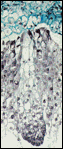
Embryos inside cultivated caryopses had been
successfully grown from initial transient stage to completely formed embryo
with plumula, radicle
closed in coleorhiza, and scutellum
(Figure 2, B). In rare cases embryos were of irregular form, with laciniate extension of scutellum
(Figure 2, C). Well-developed embryos from cultivated caryopses were isolated
and transplanted with scutellum down to the nutrient
medium for germination. The ability to germinate and to form plantlets
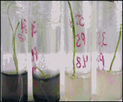
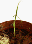
confirmed the viability of embryos from caryopses grown in vitro (Figure 3).
Sometimes embryos started to germinate straight inside cultivated caryopses on
NBM medium (Figure 2, D, E). Genotype affected the frequency of caryopses with
developed embryos (Table 3).
Table 3. Embryo formation in culture of maize
caryopses
|
Genotype |
Number of caryopses cultivated |
Frequency of caryopses (%) with developed
embryos |
||
|
in total |
of regular structure |
viable |
||
|
DK366 |
149 |
11,4 |
8,1 |
7,3 |
|
A22xDK307 |
98 |
1,0 |
1,0 |
0 |
In inbred DK366 11,4% of caryopses had the formed
embryos. 70,6% of them was of regular structure, 64,3% was viable and produced
normal green seedlings with good roots. In hybrid A22xDK307 an embryo, not
viable, has been formed only in one caryopsis. In DK366 embryos by the 20th
day in culture were of 1,76�0,39 mm long, by the 50th day – of
2,06�0,39 mm long. Significant differences in frequency of caryopses with
developed embryos at the 20th and the 50th days in
culture were not found. It may be assumed that the development of embryos takes
place for 20 days in culture, latter they only slightly enlarge.
Thus, experimental data confirm the possibility of
embryo development s in culture of isolated maize caryopses and represent the
production of viable embryos germinating in regularly developed green
plantlets. Genotype significantly affected both growth and development of
caryopses in culture, especially starch accumulation in endosperm and covers
and a frequency of developed embryos. Embryo development and caryopsis growth
were finished independently of genotype by the 20th day in culture.
Soon after this term embryos should be isolated out of caryopses for
germination and obtaining plantlets.
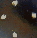
A B
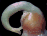
Figure 1. Maize
kernels at the moment of explantation. Caryopses (A)
and an embryo (B) at the 5th-7th day after pollination.
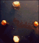

A
B

C
D
E
Figure 2. Maize kernels and embryos at the 20th day in culture. A – cultivated caryopses; B –D - embryos after isolation out of cultivated caryopses, B – embryos of regular structure; C – an embryo of irregular form, D – an embryo began to germinate inside a caryopsis; E – a caryopsis with a germinating embryo on NBM medium;
A
B
Figure 3. Germination of embryos
developed in cultivated caryopses, A – plantlets
on modified MS medium, B – a plantlet in soil.