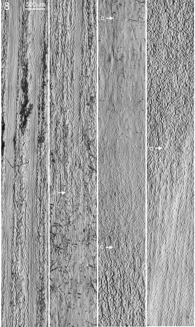
In the study of cell type, shape and size along a developing maize stem (Zea mays L., Ohio43/KYS, field grown material), epidermal replicas were obtained by applying a thin layer of clear nail enamel (New York ColorTM 138B) coating on the surface of maize stem and leaf sheath. After allowing the nail enamel to dry, the coating was carefully peeled off from the surface of the specimen and floated on a drop of water on a glass slide. The slide was then heated slightly to flatten the replica and then water was carefully withdrawn. It is important to float the replica with its cell-replicating surface facing away from the microscope slide. Optical microscopy was performed on an Olympus BX51 upright microscope equipped with an Edge dynamic oblique illumination condenser (Boyde et al., Scanning 23, 84, 2001) and an Olympus DP11 digital camera. Figures 1, 2, 3 and 4 show replica images obtained from maize stem surface (c-d region) by oblique illumination at 0o, 90o, 180o and 270 o respectively. In comparing with conventional trans-illuminated wild-field image (Figure 5), the oblique illumination provides significantly better contrast. As a reference, Figure 7 shows the surface and longitudinal section of a maize stem; the nodal region is subdivided into a-b, b-c, c-d regions. The region above "a" bears a surface typical of leaf sheath while the region below "d" consists of a surface typical of internodes (Figure 8). Our microscopy results show complex variations in the arrangement and distribution of cell types from internodes to node to leaf sheath (Figure 8). The epidermal cells on the surface of internodes are aligned in files with rows of stomata. When approaching the nodal region (Figure 8, d-b region), the stomata (S) become less organized and intermixed with other epidermal cells (c-d region, Fig. 4), then at the b-c region, stomata are completely absent. The a-b region develops a significant number of hair cells, otherwise, the arrangement of epidermal cells remain similar to the b-c region. Above the "a" level, the arrangement of epidermal cells is basically the type found on leaf sheath, with long files of silica cells (Si) demarcating the position of the vascular bundles (Figure 6) (PC Cheng et al., In: Modern microscopy, Eds. P Duke, A Michette, Plenum, 87-117, 1990). Stomata are arranged in between the files of silica cells.
Return to the MNL 77 On-Line Index
Return to the Maize Newsletter Index
Return to the MaizeGDB Homepage