Two-photon fluorescence, SHG and THG imaging was performed using a Spectra-Physics Millennia IR (1064 nm) pumped Cr-forsterite laser operating at 1230mn (110MHz, 130fs) or a Coherent Verdi pumped Spectra-Physics Tsunami mode-locked Ti-sapphire laser operated at 780mn with 100fs pulse at 82MHz.. A modified Olympus BX microscope equipped with a confocal scanning unit (Olympus Fluoview FVX) was used. A TE-cooled CCD equipped SpectraPro-150 spectrometer was used. Spectro-microscopy was performed in transmission mode by using a computer-controlled scanning stage.
Similar to the two-photon fluorescence, second-harmonic generation (SHG), which is a X(2) second-order non-linear process, provides strong nonlinear signals but occurs only in noncentro-symmetric media. SHG process can thus be used to image bio-interface and orderly arranged structure, where centro-symmetry is broken. In SHG, only virtual state transition is involved, thus no photo-damage and bleaching from the process results. With a square dependence on the incident illumination intensity, the second-harmonic generation process also provides optical sectioning resolution as the two-photon fluorescence process if the same excitation wavelengths are used. There are a number of maize structures capable of generating strong SHG signals, these include cell walls (Fig. 1), starch granules, silica cells and possibly stacked membrane structures such as grana in the chloroplast. Since they behave similarly to photonic crystals, we suggest the use of "bio-photonic crystals" to describe these types of structures. In fact, a piece of potato tuber is capable of producing a strong frequency-doubled beam similar to those non-linear crystals used in the photonic industry.
Third-harmonic (THG) is an X(3) third-order non-linear process. Like SHG, it involves only a virtual state, makes no energy deposition during the conversion process, and thus no photo-damage is expected. Due to the large refractive index difference between the fundamental and THG light and the positive dispersion in biological specimens, effective THG generation occurs only in thin layer or on the interface. This coherent length effect provides the THG process an excellent axial resolution in detecting the surface of cellular organelles, membranes and cell walls.
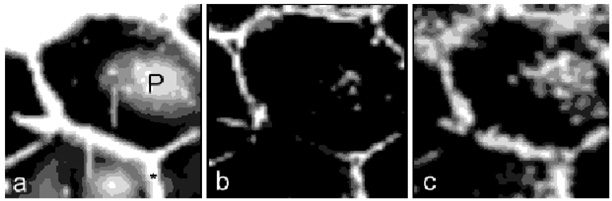
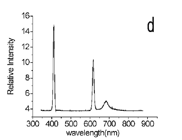
Simultaneously detecting the multi-photo fluorescence, SHG and THG provide a new way of studying biological specimens. This type of multi-modality nonlinear microscopy allows co-localization of biologically significant fluorescence signals with structural information corresponding to SHG and THG signals. Figure 1 shows a set of images (THG, SHG and red fluorescence) and a selected spectrum obtained from the parenchyma cells of the maize stem. Recording the spectrum of each pixel in an image ensures positive identification of the origin of signals.
Figure 1. Multi-modality of maize parenchyma cell showing (a) THG, (b) SHG and (c) two-photon excited fluorescence. The bright patch (P) in (a) is the transverse wall of parenchyma cell. (d) Selected spectrum taken from parenchyma cell; the excitation wavelength was 1230nm, the peak at 680nm is two-photon excited autofluorescent, the 615nm and 410nm peaks corresponding the SHG and THG signals.
Return to the MNL 76 On-Line Index
Return to the Maize Newsletter Index
Return to the MaizeGDB Homepage