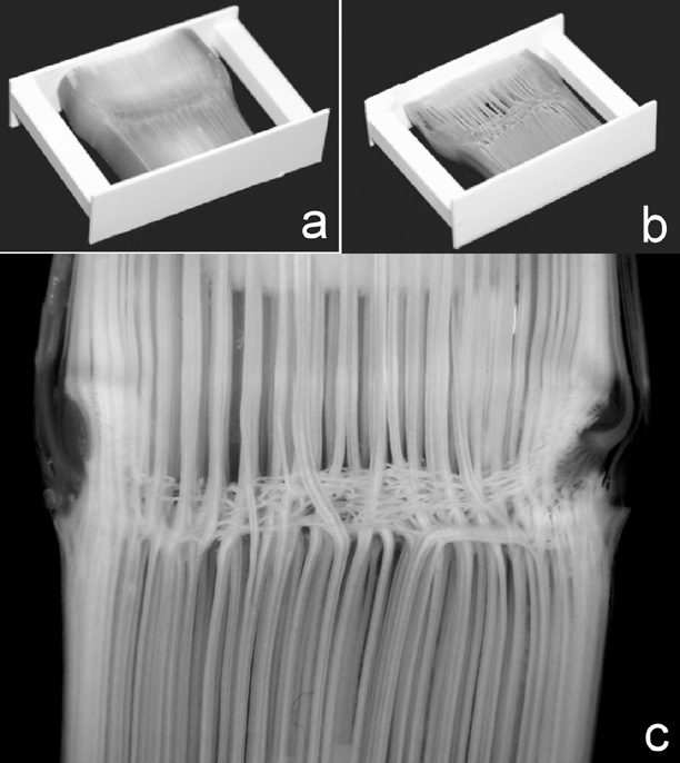
In order to preserve the vascular architecture, the stem segments when wet, were pre-glued by cyanoacrylate glue in a polystyrene frame prior to enzymatic digestion (Fig. 1-a). This prevents the vascular bundles from collapsing after the removal of parenchyma cells (Fig. 1-b). During the digestion process, tissue fragments were washed away by flushing the specimen gently with a Pasteur pipette. Figure 1-c shows the node region of Ohio43/KYS after digestion. Note the parallel bundles running through the node with branches before entering the node from below.
Figure
1. Undigested (a) and partially digested (b) stem sections glued into
polystyrene frames. (c) Digested nodal region, note the straight through
vasculatures.
Return to the MNL 76 On-Line Index
Return to the Maize Newsletter Index
Return to the MaizeGDB Homepage