Viçosa, Brazil
Universidade Federal de Viçosa
Maize root tip cell cycle synchronization
-- Carvalho, CR, Saraiva, LS, Otoni, WC
Accumulation of the cells in metaphase implies the use of some cytogenetic
strategies. The well-known spindle mitotic inhibitors action, on normal
meristematic cycle treatment, leads to metaphasic chromosome numbers appropriated
for several cytogenetic approaches. However, this chromosome amount is
not suitable for running flow cytometry analysis. This way, chromosome
accumulation procedures, previously to the metaphase blocking, must be
improved by means of an additional synchronization step. Treatments were
optimized in our laboratory to synchronize meristematic root tip cell cycles
of germinating maize seeds (test line L-869). Seeds were germinated in
Petri dishes containing a film of distilled water, and incubated at 29
C in the dark. Seedlings (1.5 to 2 cm root length) were carefully transferred
to a plastic mesh adapted inside a 6 cm diameter plastic vessels containing
100 ml of either 0, 1, 2, 4, and 6 µM hydroxyurea (HU). After two
cycles, approximately 18 h, the roots were recovered by washing for 15
min (running tap water) and incubated in distilled water under the same
conditions as before. Thereafter, samples of root tips were taken from
0 to 10 h, at 1 h intervals, and fixed in a fresh ice-cold methanol:acetic
acid solution (3:1), and kept in a freezer for at least 24 h. Next, root
tips were excised at 0.1 cm and macerated with 200 µM of freshly
prepared Flaxzyme (NOVO) enzymatic solution plus 1.6 ml distilled water,
and incubated at 35 C for 2h 30 min. The macerated cells were dissociated
in a clean slide with a freshly fixative solution, air-dried and stained
with a Giemsa solution. Meiotic figures were photomicrographed and digitized
directly by a microscope-coupled CCD video camera to a computer. Under
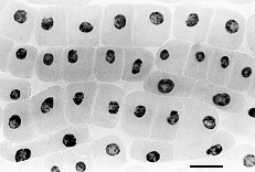
the experimental conditions, 2 µM HU enabled higher cell synchronization
indexes (Figure 1) as compared to higher HU concentrations. By using only
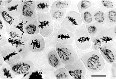
the synchronization step, at 2 µM HU, combined with 6-7 h recovering
time, an average of 28% of metaphasic cells (Figure 2) was obtained in
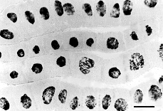
comparison to 0 µM HU control treatment (Figure 3), that normally
displayed less than 1% of metaphasic cells. It was also noted that at 6
µM HU the cells remained in interphase, therefore losing the reversibility.
This technique proved to be reproducible, being applied not only to cytogenetic
and cytometric purposes, but to a wide range of cell cycle studies.
Figure
1. Highly synchronized interphasic maize root tip cells after 18 h
at 2 µM HU. Bar = 20 µm.
Figure
2. Highly synchronized metaphasic maize root tip cells obtained after
2 µM HU treatment and 6-7 h recovering time without HU. Bar = 20
µm.
Figure
3. Typical pattern of maize root tip cell cycle from control treatment
(0 µM HU). Bar = 20 µm.
Please Note: Notes submitted to the Maize Genetics Cooperation Newsletter may be cited only with consent of the authors.
Return to the MNL 76 On-Line Index
Return to the Maize Newsletter Index
Return to the MaizeGDB Homepage