The role of microfilaments in cell wall formation through the progression of microsporogenesis was explored using two maize male sterile mutants. This comparative study utilized ms2, a postmeiotic mutant of maize in which developing microspores are deficient in cell wall synthesis, and po, a mutant that develops a normal cell wall despite the occurrence of a number of postmeiotic disturbances. The first mutational abnormalities are observed to initiate at the same stage, in the tetrad stage after meiosis II, in both of these mutants. A wild-type strain was also studied as a control.
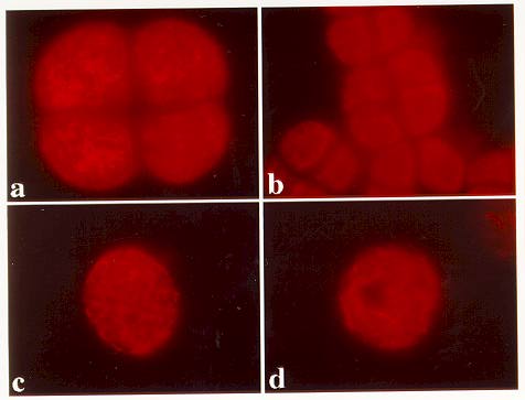
When compared to the microfilament array of similarly staged wild-type microspores (Figure 1a), the ms2 mutants do not display a reticulate staining pattern that is consistent throughout the cytoplasm. Instead, microfilaments combine to form thick cables that display little contact with the membrane (Figure 1b), except for a small number of fine microfilaments that branch off actin cables, which are oriented parallel with the membrane. After release from the tetrad, microspores never form an ordered cortical staining pattern typical of wild-type microspores of the same stage, but instead show a more random cytoplasmic array that displays plasma membrane contact (Figure 1c). As development proceeds, the microfilaments become fewer in number as they condense into very thick microfilament cables, which extend randomly throughout the cytoplasm (Figure 1d).
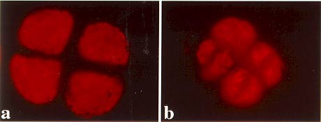
Abnormalities in microfilament arrays are observed in the po mutant only after the tetrad stage, upon initiation of post-meiotic divisions. Following tetrad formation, the microfilament array appears quite normal, and exhibits formation of small actin patches visible in most cells (Figure 2a). Shortly after this stage, coinciding with the onset of abnormal post-meiotic divisions, the microfilament array appears to constrict or shrink within the cells, lending an appearance of a more disorganized microfilament array (Figure 2b). After microspore release from the tetrad, the cell wall develops to a normal thickness, despite the absence of an organized microfilament array and an intact nucleus.
An interesting correlation between the absence of cell wall formation, and an abnormal microfilament array existed during development of ms2 mutant microspores. At similar stages wild-type and po microfilament arrays are observed to extend throughout the cytoplasm, and to contact the cell membrane at many points. Cortical actin patches were observed in wild-type and po tetrads, but were not present in ms2 tetrads. Similar structures have been found in yeast, where the location of these actin patches has been shown to correlate with new cell wall synthesis (Gabriel and Kopecka, Microbiology 141:891-899, 1995; Mulholland et al., J. Cell Biol. 125:381-391, 1994). If the actin patches observed in wild-type and po represent analogous structures to those observed in yeast, the early tetrad stage could represent the time period critical to the initiation of normal cell wall formation in maize microspores.
Figure 1. Wild type and ms2 microspores stained to visualize microfilament distribution. a) a wild type tetrad showing a normal reticulate staining pattern; b) ms2 mutant microspores in which microfilaments join to form thick cables; c) ms2 microspore after tetrad release; d) ms2 microspore after a longer period of development.
Figure 2. polymitotic microfilament distribution. a) microfilaments show a normal distribution before the onset of post-meiotic divisions; b) microfilaments become more disorganized as cells undergo abnormal divisions.
Return to the MNL 72 On-Line Index
Return to the Maize Newsletter Index
Return to the MaizeGDB Homepage