We have reported on the inducible heat shock (hs) genes of maize and the modulated transcriptional activity of some, but not all, of these genes during microsporogenesis and gametogenesis (Atkinson et al., Dev. Genetics 14:15-26, 1993; Bouchard et al., Maydica 38:135-144, 1993). Brothers et al. (MNL 67:73-74, 1993) reported on the distribution in maize root tip cells of polyclonal antibodies raised to the 18 kDa family of HSPs. The antibodies were raised against protein isolated from hs root tips.
To extend our study, we undertook to locate the sites in root tip cells of mRNA for the 18 kDa family of HSPs. We report below the hybridization procedures employed on root tip sections for the antisense RNA (and sense controls) to the mRNAs containing the open reading frame (ORF) from the family of 18 kDa HSPs.
Preparation of probes. Several of the 18 kDa maize heat shock protein (HSP) genes (Goping et al., Plant Mol. Biol. 16:699-711, 1991) were cloned into pBluescript II Sk- vector containing T7 and T3 RNA polymerase promotors. Template DNA of scMHSP 18-9-2 (a 342 bp fragment containing the ORF) was linearized prior to RNA synthesis and both sense (control) and antisense transcripts were synthesized. The scMHSP 18-9-2 fragment was cut with XbaI + T7 polymerase for the sense strand and PstI +T3 polymerase for the antisense strand.
After restriction enzyme digestion, the template was purified by phenol/chloroform extraction according to standard procedures and dissolved in DEPC-H2O. Transcripts were produced from 1 µl of template DNA and 6-8 µl of digoxigenein (DIG) RNA labelling mixture (Bohringer-Mannheim) [60-80 mM of all nucleotides]. The concentration of the sense and antisense probes was 10 ng per 1 µl of hybrization solution (buffer prepared according to Langdale (In Freeling and Walbot, eds., The Maize Handbook, pages 165-180, Springer-Verlag, 1993). We used 15 µl per slide (for a 22 mm2 cover slip).
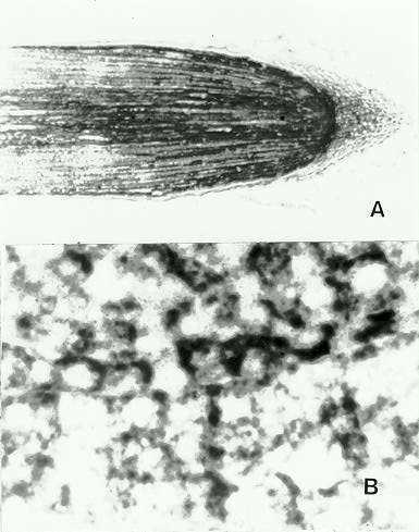
Hybridization procedures. 10 µm longitudinal sections (l/s) of FAA-fixed, paraffin embedded root tips from heat shocked and control seedlings (4-5 day) were tested for in situ hybridization of hs mRNA using a modified procedure from Langdale (1993). Depending upon the length of treatment with the substrate solution (currently 10 h) duration and temperature of post hybridization washes, obvious and consistent hybridization (dark blue-purple staining) was observed with the antisense mRNA of the 18-9-2 clone (Fig. 1A). Considerably lighter or no staining was detected with the sense probes on hs root tip sections and with both sense and antisense probes on sections of control root tips.
The hybridization was distributed unevenly throughout the 1cm portion of the root with the greatest intensity towards the meristem (Fig. 1A). Cytologically the staining was restricted to the cytoplasm with dense accumulations and with many dispersed punctate sites (Fig. 1B). Attempts to quantify the different responses of different tissues of the root are continuing as are comparative studies with probes derived from other members of the 18 kDa family of HSPs.
Figure
1A: Near-median l/s through maize root tip documenting the typical
staining response to the antisense DIG probe (X25). Figure
B: View of response of cells from the region of the root tip meristem
and the root cap to antisense DIG probe (X900).
Return to the MNL 68 On-Line Index
Return to the Maize Newsletter Index
Return to the MaizeGDB Homepage