The dominant Hsf1-O (Hairy-sheath frayed) mutant of maize has been shown to retard the transition of juvenile cells to adult. One of the most obvious Hsf1-O phenotypes is the transformation of leaf blade cells into sheath, resulting in prong-like projections of sheath tissue along the edges of the blade (Bertrand-Garcia and Freeling, Am. J. Bot. 78:747, 1991).
In normal maize leaf development, as basipetal cell differentiation along the leaf occurs, a ligule develops perpendicular to the plane of the leaf separating blade tissue from sheath. Likewise, prongs may have their own ligule growth, at the boundary between sheath and blade, so that a single leaf will have more than one ligule. The development of the prong is spatially separate from that of the normal blade-sheath boundary. This spatial separation of ligular forming regions in Hsf1-O mutants presents an excellent model in which to study ligule development and to further understand the nature of the proposed signal for the initiation of ligule growth (Becraft and Freeling, Plant Cell 3:801, 1991).
Plants heterozygous for Hsf1-O in a B73 inbred background were observed for prong formation. These plants show the development of prongs, as early as leaf numbers 5-7. In young seedlings, prong initiation can be seen occurring distally, within 1.0cm of the normal sheath-blade boundary. Leaves 5-7 showing one or more prongs were examined under a dissecting microscope to determine the approximate state of prong differentiation. Twenty prong specimens in the earliest stages of development (1.0cm from the sheath-blade ligule) were removed from the leaf along with the normal ligular region. These specimens were fixed, sputter coated with gold and examined by scanning electron microscopy.
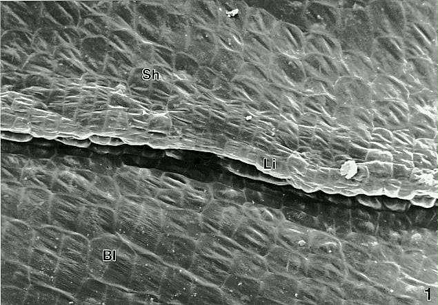
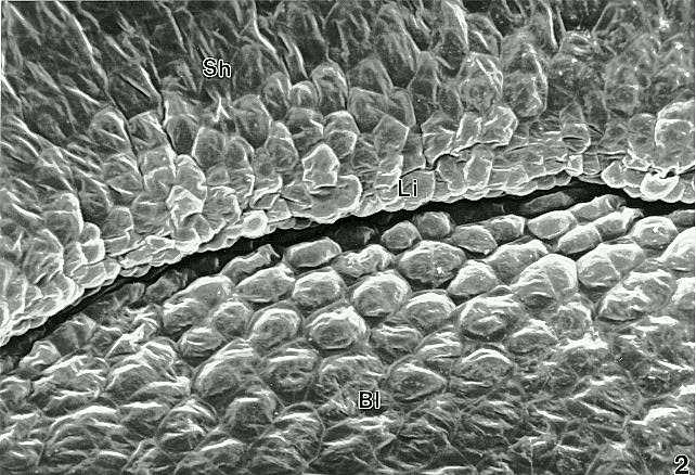
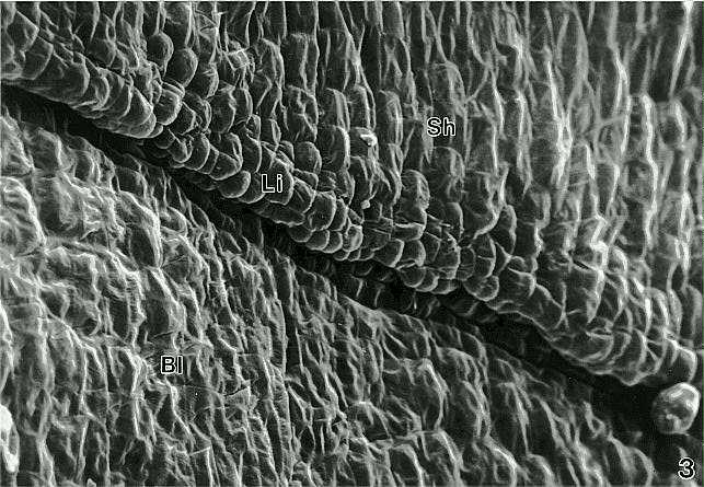
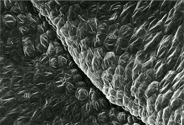
Comparison of ligule initiation at the normal sheath-blade boundary and within the prong of the same leaf shows the simultaneous initiation of the ligule at both sites (Figs. 1-4). This finding would support a model of a single ligule signal that originates at the tip of the leaf and diffuses basipetally. Only cells that are competent (at the appropriate developmental stage) to receive and respond to the signal then proceed on to initiate ligule development. If more than one signal existed or if the ligule signal was continuously produced we would expect cells in the prong region to respond first and for ligules in the prong region to be more developed. These results further suggest that cells in the prong, although chronologically older than those in the normal blade-sheath region, are retarded in their development and remain competent to receive the ligule signal. This model affirms previous investigations which suggested that Hsf1-O is a heterochronic mutant.
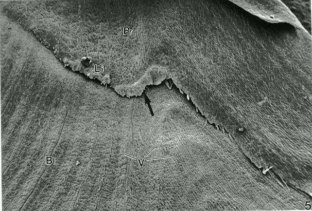
Although this study concludes that normal and prong ligule development are initiated simultaneously, it is interesting to note that prong ligules do not always develop fully. It is more likely to observe several stages of maturation along the prong, the most advanced at the center of the transformed region, where the veins of the leaf are seen to break their parallel lines and bend into the prong (Fig. 5). This appears to be the point of origin for both the prong and ligule development along the rim of the blade.
The sporadic occurrence of ligule in some prongs and not in others, and the variable maturation of the ligule in prongs, may occur (1) because of temporal separation, the competent cells in the prong may be reaching a state in which they can no longer respond normally to the signal, or (2) because fewer cells in the region can respond. Further studies using this mutant will address questions regarding the level of determination in those cells competent to receive the ligule signal.
Figure 1. Early ligule (Li) development at the normal boundary between sheath (Sh) and blade (Bl) on Hsf1-O plant, leaf number six. (x460)
Figure 2 Ligule development at prong area from leaf shown in Figure 1. (x460)
Figure 3 Example of ligule development at the normal sheath-blade boundary in Hsf1-O. Figure shows leaf number twelve. (x460)
Figure 4 Prong region with ligule development, from leaf shown in Figure 3. (x460)
Figure
5 Prong (Pr) growth along the edge of the blade. Note how the typically
perpendicular veins (V) "bend" inwards from the blade (Bl) towards the
center of the ligule (Li), where it is most developed. This region (arrow)
is the point of ligule initiation in the prong.
Return to the MNL 67 On-Line Index
Return to the Maize Newsletter Index
Return to the MaizeGDB Homepage