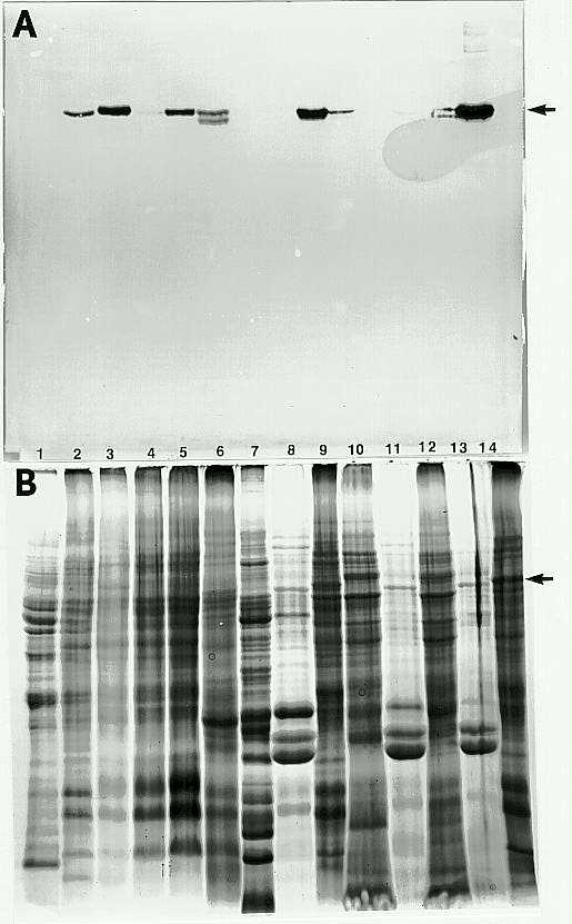
Soluble and total protein were extracted from a variety of plant organs and parts in order to quantitatively determine enzyme activity and the amount of antigen reacting with anti-beta-glucosidase sera in extracts of each of these organs and parts. The plant organs and parts used were: pollen, male spikelet, rachis, silk, husk, ear axis, ovule, mature leaf, germ and endosperm dissected from seeds on day 0, 3, and 5 after germination (DAG), and coleoptile. Figure 1 shows that the highest enzyme activity was measured in the coleoptile, primary root, husk, and ovules. Low levels of enzyme activity were measured in the silk, spikelet, mature leaves, and germ (3 DAG and 5 DAG), while no activity was detected in extracts of whole mature kernels, pollen, rachis, ear axis, and endosperm. The immunoblotting results (Figure 2) confirm those from spectrophotometric assays in that only the organs and parts that have detectable enzyme activity have a 60kD (arrow) immunoreactive polypeptide (beta-glucosidase monomer).
The overall results from studies on organ specificity of beta-glucosidase expression show that the enzyme is exclusively found in actively growing plant parts (e.g., primordial leaves, shoot apex, mesocotyl, primary roots) of young seedlings or female reproductive organs (e.g., ovule, silk, and husk). Plant parts that are terminally differentiated and matured (e.g., endosperm, ear axis, and rachis) are either devoid of enzyme activity or the enzyme is not extractable with aqueous buffers even after SDS has been added to a 0.5% final concentration. Moreover, the presence of SDS in the extraction buffer did not affect the amount of activity extracted, except in the case of roots, where SDS helped extract additional activity. These results suggest that beta-glucosidase is a key enzyme with critical role(s) in normal plant growth and development. Our laboratory could not find any evidence for cyanogenesis in maize coleoptile extracts using either maize or sorghum beta-glucosidase as the enzyme source. Interestingly, the coleoptile (coleoptile proper, mesocotyl, primordial leaves, and shoot apex) is the richest source of the maize enzyme, but it is not a cyanogenic plant part, suggesting that the reactions catalyzed by the enzyme in young shoots do not involve cyanogenic glucosides as substrates.
Figure 1. Tissue-specificity of maize beta-glucosidase expression. The enzyme was extracted from freeze-dried powder of each plant part with 0.1M Tris-HCl, pH 8.0, containing minus or plus 1% SDS, and the extracts were assayed for activity after 100X dilution with the standard enzyme assay buffer. Note that the highest total activity was measured in the coleoptile extract followed by root, husk, and ovule extracts, respectively.
Figure 2. Tissue-specificity of maize beta-glucosidase expression. The extracts used for activity assays (refer to Figure 1) were subjected to SDS-PAGE, blotted onto nitrocellulose, and probed with anti-beta-glucosidaseserum. Lane 1, pollen; 2, silk; 3, husk; 4, ear axis; 5, ovule; 6, leaf; 7, germ at day 0; 8, endosperm at day 0; 9, primary root; 10, germ at day 3; 11, endosperm at day 3; 12, germ at day 5; 13, endosperm at day 5; and 14, coleoptile.
Return to the MNL 67 On-Line Index
Return to the Maize Newsletter Index
Return to the MaizeGDB Homepage