ESALQ, Universidade de São Paulo
Mitotic instability in callus cultures of inbred lines adapted to tropical regions
--Margarida L. R. de Aguiar-Perecin and Antonio Fluminhan, Jr.
A procedure to investigate aspects of mitotic instability occurring in callus culture is presented in this report. We analyzed cultures initiated from F2 immature embryos derived from the cross between two sister lines designated 300-14-1315 (S7) and 300-14-13213 (S8), which have Type II (friable and embryogenic) culture response. These lines are derived from a flint variety (Jac Duro, Sementes Agroceres) developed in Brazil, and are homozygous for C-bands correspondent to K6L2/K6L3, K7S, K7L, K8L (L1 or L2, not determined), and K9S (references in MNL 62:100, 1988).
The cultures were incubated on MS medium containing 2mg/L 2,4-D and 20mg/L casein hydrolisate (Fluminhan and Aguiar-Perecin, Ciencia e Cultura Suppl. 43:760-761, 1991). Mitotic analysis was carried out on meristematic cells of proembryoids taken from the callus 5 days after its transferrence to fresh medium, fixed in 3:1 alcohol:glacial acetic acid and stained in Feulgen. C-banding was also employed. Proembryoids were squashed in 45% acetic acid after incubation in 2.5% pectinase. For metaphase preparation a pretreatment with 0.002M hydroxiquinoline was carried out.
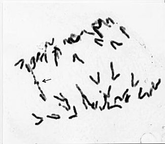
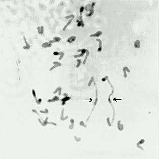
The analysis of anaphase bridges showed two types of figures: i) bridges which appear to be the result of delayed segregation of sister chromatids (Fig. 1); ii) bridges (with and without bands) apparently originating from a breakage-fusion-bridge cycle initiated by broken chromosomes (Fig. 2). These observations are consistent with the interpretation that the chromosome aberrations, usually involving knobbed chromosomes, observed in regenerants are the result of chromosome breakage occurring in culture (Phillips, RL et al., Corn and Corn Improvement, pp. 345-388, 1989).
Figure 1. Feulgen stained anaphase showing one bridge apparently derived from delayed segregation of sister chromatids.
Figure 2. C-banded anaphase with two bridges involving a banded chromosome arm. They appear to be originated from previously broken chromosomes.
Table 1. Frequency of mitotic anaphases with bridges in 3 cell lines.*
| Anaphases | ||||
| Cell line designation | Weeks in culture | Normal | 1 bridge | 2 bridges |
| H | 26 | 25 | - | - |
| J | 26 | 103 | 4 | - |
| H | 39 | 20 | - | 1 |
| H | 43 | 140 | 6 | 1 |
| 57 | 53 | 141 | 4 | 3 |
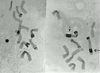
Figure 3. C-banded metaphase showing enlarged bands at K7L.
We analyzed the frequency of bridges in some cell lines as shown in
Table 1. Anaphases with 1 and 2 bridges were observed in these samples.
A preliminary observation of metaphases showed clear alterations of the
structure of chromosome 7, such as the presence of 3 bands, K7S on a subterminal
chromosome position and amplification of K7L (Fig. 3). These aspects suggest
that in most cases this is the chromosome involved in the formation of
bridges. One interesting aspect concerning the chromosomes with enlarged
bands is that their pattern is not altered. If this amplification is an
initial event that leads to the delay of chromatid segregation, it is an
interesting point for further investigation. A complete report on the frequency
of anaphase bridges in cultures of endogamic lines with different knob
compositions is in preparation.
Return to the MNL 66 On-Line Index
Return to the Maize Newsletter Index
Return to the MaizeGDB Homepage