State University of New York
ROCHESTER, NEW YORK
University of Rochester
--Ping-chin Cheng and Hyo-gun Kim
The ability to absorb soluble silica from the soil, and subsequently translocate and deposit it in various parts of the plant (Lanning and Linko, J. Agric. Fd. Chem. 9:463, 1961) in the form of SiO2.nH20, has been observed in many species, but particularly in grasses, for decades (Jones and Handrek, Adv. Agron. 19:107,1967). It was reported that silicon in living rice plants is present in three basic forms: 1) insoluble silica (90%), 2) silicate ions (0.5-8%) and 3) colloidal silicic acid (0-3.3%). Furthermore, the mobility of silicon in rice plants is poor. In other words, reutilization of silicon, once deposited in plant tissue, is very unlikely to occur (Yoshida et al., Soil Sci. Plant Nutrition 8(3):15, 1962). The insoluble silica in plant tissue is generally a clear, colorless and isotropic deposit with an index of refraction of 1.42 to 1.44 (Jones and Handreck, 1967). X-ray diffraction studies revealed an amorphous pattern similar to opals. Because of these physical properties, silica deposits in plants are believed to be similar to opal minerals; hence, they are frequently referred to as being biogenic opals.
Jones and Handreck (1967) pointed out that the deposition of silica in plant tissue cannot be readily studied under the microscope without special treatment of the plant material. Traditionally, six methods are used for the microscopic determination of silica deposits in plants. They are: 1) formation of sodium silicofluoride crystals by treating the tissue with NaCl and HF; 2) wet ashing; 3) dry ashing (spodogram); 4) staining with basic fuchsin, safranin-phenol and malachite green; 5) hydrofluoric acid etching of plastic-embedded tissue (Yoshida et al., 1962); and 6) mounting the tissue in a high refractive index medium. Recently, Dayanandan et al. (Amer. J. Bot. 70:1079, 1983) reported the use of histochemical reactions based on the reactivity of the silanol (SiOH) group of silica and the use of polarized microscopy to detect a special type of silica body (phytoliths) in plants. The methods listed above involve either harsh chemical treatment or high temperature ashing of the tissue. In addition, x-ray microanalysis (EDS) has been used extensively in the detection and mapping of mineral deposits including silica. The EDS method works well on detecting silica on the surface of bulk specimens, sectioned or fractured sample. However, the EDS technique is restricted to the very surface layer of the specimen. Therefore, detection of silica in deeper layers of tissue is not possible. Due to the deeper penetration, back scattered electron (BSE) imaging in a scanning electron microscope has also been used to image silica cells.
Due to the properties of x-rays and the availability of various high intensity x-ray sources in recent years, x-ray microscopy becomes a useful tool in the study of silica deposits in plants. We report here the use of x-ray contact microradiography using a pulsed x-ray source for the study of the silica deposition in the leaf blade of corn. This method not only allows the examination of silica deposition in dry specimens, but most importantly, it makes imaging of living specimens possible (X-ray Microscopy, eds. P. C. Cheng and G. J. Jan, Springer-Verlag, 1987). In contrast to conventional electron beam microanalysis (EDS), which is restricted to the very surface layer of the sample, x-ray imaging allows the study of silica deposits deep in the tissue.
The maize used in this study was greenhouse grown plants of Golden Beauty. Either fresh leaf blade or aldehyde-fixed (Cheng et al., Natl. Sci. Counc. Monthly, ROC 7:1001, 1979) tissue were used. Fixed leaf blades were dehydrated in acetone and critical point dried in CO2. For controls, fixed leaf blades were treated in a 5% HF solution for 48 hours to remove the silica deposits, and then washed, dehydrated and critical point dried. The silica composition of the deposits was confirmed by using energy dispersion spectroscopy (EDS) in a scanning electron microscope.
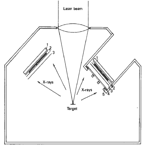
A laser-produced plasma x-ray source was used in this study. A frequency tripled high intensity Nd:glass laser beam (Glass Development Laser at the Laboratory for Laser Energetics of the University of Rochester) was focused onto a Mo thin target (Figure 1). A one nanosecond pulse was delivered to the target, which generates a high temperature (107K)plasma. Laser output ranging from 20 to 100J of ultraviolet radiation was used in this experiment. The x-ray source size was on the order of 100µm and the specimen was placed approximately 25cm away from the source.
Figure 1. Diagramatic representation of the x-ray contact microradiography set-ups. Left: for the imaging of dehydrated specimen. Right: for the imaging of fresh sample. 1: film plate, 2: two sheets of Mylar film with specimen sandwiched between them, 3: Al filter, 4, 2um thick Mylar vacuum window, 5 1mmx1mm stainless screen. The target is in vacuum.
The experimental set-ups are shown in Figure 1. The x-ray contact imagings of dehydrated specimens were done in vacuum, and fresh tissues were imaged under atmosphere pressure by using a 2µm thick Mylar film as the vacuum window. An Al filter (two 250nm thick Al layers evaporated on both surfaces of a 2µm thick Mylar film) was used to block the UV and visible radiation emitted from the high temperature laser-produced plasma. The samples were sandwiched between two Mylar foils, and held in close contact with the photographic plate. The x-ray contact microradiographs were recorded on Agfa-Gavaert holographic plates (emulsion 8E75); the plate has a resolution of the order of 2000 lines/mm. The holographic plates were developed in Kodak D-19 for 10 min. The contact images were magnified by using either a macrophotography set-up or a compound microscope (Cheng et al., In: Modern Microscopy, eds. P. Duke and A. Michette, Plenum Press, 1989).
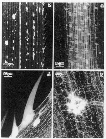
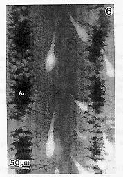
The dense silica deposits in the leaf blade can be easily detected by x-ray contact microradiography (Figure 2). As revealed by the x-ray imaging technique, silica is initially deposited in specialized dumbbell-shaped silica cells (Figure 3) which are in close association with the vasculature, excluding the main vein. In the mature leaf, it is common to find that silica deposits in the hairs (Figure 4), the epidermal cells surrounding the base of the epidermal hairs (Figure 5), and in selected patches of epidermal cells (Figure 2). Figure 6 shows the x-ray image of silica deposition in a fresh leaf blade.
Figs. 2-5. Figure 2. X-ray contact image of a leaf blade of maize. Figure 3. Dumb-bell-shaped silica cells (Si). Figure 4. Siliceous hairs. Figure 5. Siliceous epidermal cells at the base of a hair (Hr).
Figure 6. Siliceous hairs in a fresh leaf blade. Ar: air space in leaf blade.
The use of x-ray contact microradiography in conjunction with a high intensity x-ray source offers a new technique to study the silica deposition in fresh tissue. Technical simplicity is not the only advantage that x-ray contact microradiography can offer over other methods, the most important of which is that the technique allows the study of living specimens with a very large "field of view". At the present time, we can image a leaf area as large as 4x4cm, and the limit of the field of view is only restricted by the size of the photographic plate. Studies have shown that there is a correlation between a high concentration of silica deposits and insect resistance in many plant species. For instance, Ponnaiya (Madras Univ. Jour. XXI, Sect. B., No. 2: 203, 1951) reported that the deposition of irregular silica particles in the leaf sheath of a variety of sorghum correlated with resistance to the larva of Antherigona indica. High silica content in rice has also been correlated with resistance to the Asiatic rice borer, Chilo suppressalis (Djamin and Pathak, J. Econ. Ent., 60:347, 1967). Lanning et al. (Ann. Bot. 45:549, 1980) suggested that breeding for high silica content could be a potential factor for developing insect-resistant corn varieties. Therefore, the large "field of view" offered by x-ray contact microradiography can be used to screen large areas of leaf to obtain statistically meaningful data, such as patterns of silica cell distribution, needed in breeding programs. Furthermore, the technical simplicity of x-ray contact imaging also allows the possibility of screening large numbers of samples in a short time.
This work was supported by the U.S. Department of Energy under project
DE-FC08-85DP40200, DE-A508-88DP10782 and the Biomedical Research Supporting
Grant Program, NIH (BRSG SO RR07066). Special thanks to Mr. T. V. Bieniek,
K. Liu, W. Schulze, and M. D. Wittman for their wonderful technical assistance.
Return to the MNL 63 On-Line Index
Return to the Maize Newsletter Index
Return to the MaizeGDB Homepage