--Phil Becraft and Michael Freeling
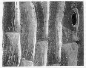
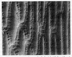
For developmental investigations it is essential to recognize the constituent cells and tissues of the particular system. Scanning electron microscopy (SEM) is proving useful for leaf developmental studies. Using SEM, we have found that the epidermis of most leaf parts has characteristic and distinctive surface features, and it has been possible to distinguish leaf tissues which otherwise appear very similar. For example, the gross morphology of sheath and midrib is nearly identical; each are thick, rigid, and have a smooth, shiny adaxial surface. In cross section, both have thick mesophyll layers with sparse chloroplasts, and vascular bundles, and are associated with prominent fibers localized toward the abaxial surface. Genotypes causing anthocyanin pigmentation in the sheath often affect the midrib as well. Thus it has been difficult to discern sheath and midrib by means other than position on the leaf. However, SEM images of the adaxial epidermis show clear distinctions between sheath and midrib. Sheath epidermal cells (Figure 1) have wispy wrinkles running longitudinally, are rectangular in shape, and have narrow cell borders. The midrib epidermal cells (Figure 2) have smooth wall surfaces, are rectangular to somewhat diamond shaped, and have a distinctive weld-like appearance at the cell borders. No other cells with these surface characteristics were found in normal maize leaves.
Figure 1. Scanning electron micrograph of adaxial sheath epidermis. 507X.
Figure 2. Scanning electron micrograph of adaxial midrib epidermis. 520X.
Several mutations exist in maize which appear to transform tissue of
one organ component into that of another organ component (e.g. Hsf1
transforms blade to sheath-- older to younger). Knotted plants (the phenotype
of Kn1 ) have patches of sheath-like leaf, often bounded by ligular
fringes, along lateral veins of the blade. SEM provided conclusive evidence
that this sheath, although located in the blade, is actually sheath rather
than midvein or any other leaf component. Thus, we have established leaf
cellular identities independent of position within the leaf.
Return to the MNL 63 On-Line Index
Return to the Maize Newsletter Index
Return to the MaizeGDB Homepage