Tissue cultures utilizing immature embryos of a field F1 red flint hybrid, 81-719 x 81-699, obtained by Francisco Babinec, were initiated in February 1982. The culture protocols were the same as mentioned above for the floury-a immature embryos.
Immature embryos of 15, 18 and 20 days after pollination were placed with their scutellar side upwards on MS medium containing 2,4-D at 2, 5 and 10 ppm. 2 ppm 2,4-D medium and 20-day embryo age was the most successful combination to promote callus formation. After 4 subcultures, 30 days each, more than 20 test tubes with 0.5 - 1.5 cm diameter callus were developed from each primary culture. After the fourth subculture, attempts were made to subculture callus tissues in medium free of 2,4-D to promote organ formation. This organ formation phase was carried out at 30/20 C and 12/12 hours photoperiod.
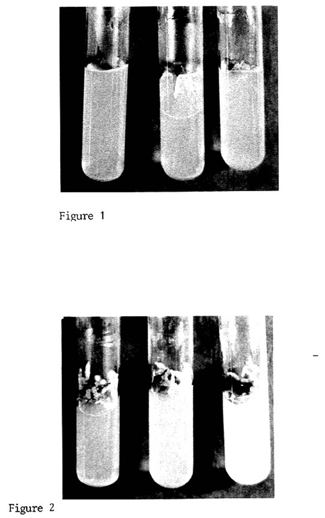
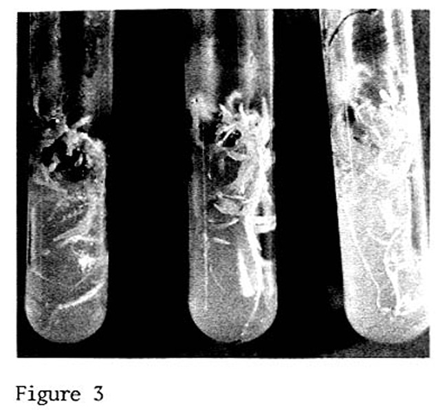
After 5 months of culturing, a calliclonal variation synthesized in three kinds of responses was observed in the cultures. Some cultures (Fig. 1) did not grow further during subsequent inoculation, and were brownish and usually necrotic after 60 days. No organized root vascular tissue was observed. A second kind of response (Fig. 2) was observed in the majority of the cultures, and was a kind of growth that appeared as a mixture of callus, agar roots and principally aerial roots. Both agar and aerial roots were hairless. A third kind of response (Fig. 3) was observed in 17% of the subcultures, and was characterized by a development of extensive roots with root hairs and few aerial roots. One of the cultures presented more than 40 adventitious roots with secondary and tertiary roots.
None of the cultures with extensive root development produced shoots at this time. Such observation is in agreement with Green and Phillips (Crop Sci. 15:417-21, 1975). Structures resembling organized scutellum were not observed in the cultures. Apparently, the root development appeared from the mass callus.
Attempts were made to characterize electrophoretically the three kinds of responses by conventional PAGE (Davis) and SDS-PAGE (Laemmli). However, the low protein content of the tissues was a difficult risk in our attempts. As much as the equivalent of 250 mg fresh weight of tissues was loaded in each acrylamide tube without obtaining successful visualization of the protein bands with Coomassie Blue R-250. However, it was evident that the SDS-PAGE patterns derived from each kind of culture were different and distinguishable. More than 25 bands were observed in the SDS-PAGE patterns of the cultures of Figure 3, while only 5 to 10 bands were observed in the SDS-PAGE patterns of the cultures of Figure 1.
Miguel Angel Rapela
Return to the MNL 57 On-Line Index
Return to the Maize Newsletter Index
Return to the MaizeGDB Homepage