With the development and refinement of plant tissue culture techniques, the prospects of chromosome manipulation at the cellular level are quite promising. Plant tissues and cells, like their animal counterparts, display more than the usual degree of genomic instability when they are removed from the stabilizing environment and plunged into the alien environment of a culture vessel. This ranges from increases in ploidy level to different degrees of aneuploidy. Given the widespread intentions to apply in vitro methods to somatic cell genetics, it is particularly important to develop a suitable banding methodology, which would aid not only in the identification of structural changes but also in the location of those points at which chromosomes break.
In the present communication, a suitable method for Giemsa banding in analysis of chromosomes under in vitro conditions is presented. It is interesting to note that all the numerical and structural changes begin very early in cultures of different explants of Zea mays cv. compositae. As a result, a number of abnormal karyotypes emerged. The revised method of Giemsa banding includes the following steps:
Pre-treatment: Callus tissues from the growing regions were pre-treated with a saturated aqueous solution of alpha-bromonaphthalene at 14-18 C for 4 hours, after placing them in sunlight for 1-2 hours.
Fixation: In 1:3 acetic-alcohol at 10 C for 4-12 hours and preserved in 70% alcohol.
Maceration: In 9:1 orcein:HCl mixture at 60 C for 5-6 min. Callus tissues were squashed in a drop of 45% acetic acid. Cover glasses were detached from the slides by immersing them in ethanol, and both slide and cover glasses were air dried.
Destaining: In 45% acetic acid for 10-15 min. Washed in distilled water for 5-10 min and air dried.
HCl treatment: With 5 N HCl at room temperature for 5-7 min. Washed in running distilled water for 15 to 20 min.
Staining: With 3% Giemsa solution diluted in 1/15 M sodium or potassium phosphate buffer at pH 6.8 for 30 seconds to 1 min. The staining process was monitored by microscopic observation of the slides at frequent intervals.
After adequate staining, the slides were rinsed in distilled water and air dried before mounting in Canada balsam.
The revised method of banding showed that fixation in acetic-alcohol plays a promoting role for the development of bands. This may be due to the disruptive lesion of chromosomal protein, which is primarily responsible for band development.
In addition, experimental conditions indicate that the ionic strength of the salt solution has some significant role in band development. Therefore, it can be inferred that the acetic alcohol and concentration of HCl might dissociate certain substantial amounts of protein from the chromosomes to which the Giemsa binds and ultimately develop a band. The mechanisms involved are yet to be clarified.
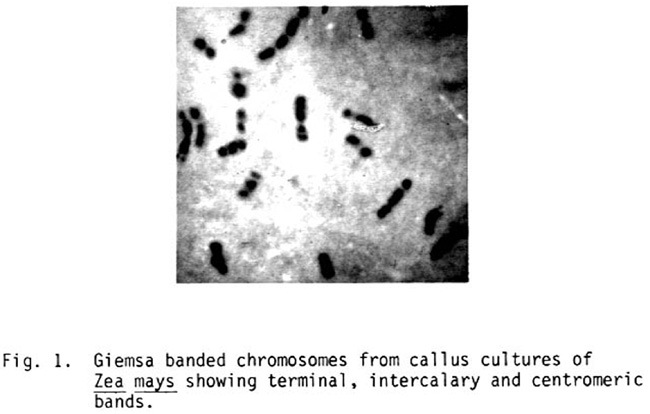
Fig. 1. Giemsa banded chromosomes from callus cultures of Zea mays showing terminal, intercalary and centromeric bands.
Most of the chromosomes showed terminal, interstitial, as well as centromeric bands (Fig. 1). Such a type of banding pattern not only helped to identify the chromosomes which have been eliminated preferentially during the culture regime, but also to detect structural changes in the chromosomes.
N. K. Paul and P. D. Ghosh
Return to the MNL 57 On-Line Index
Return to the Maize Newsletter Index
Return to the MaizeGDB Homepage