Sucrose synthetase is coded by the Sh1 locus on chromosome 9. Chourey and Nelson (Biochem. Genet. 14:1041, 1976) demonstrated that a residual sucrose synthetase activity in sh1 strains could be attributed to a protein with a slightly faster mobility on native gels. The two proteins are similar by many criteria (immunochemical, catalytic, subunit molecular weight) but show different developmental profiles (Chourey, Molec. Gen. Genet., in press, 1981). This report presents further evidence for the hypothesis (Chourey and Nelson, 1976) that the two sucrose synthetases are coded by two separate genes.
There are 3 maize strains available that are believed to be partial or complete deletions of the Sh1 gene on chromosome 9. These are the two x-ray induced mutants sh-bz-x2 and sh-bz-x3 (Mottinger, Theor. Appl. Genet. 43:190, 1973) and sh-bz-m4 (McClintock, Carnegie Yearbook 55:323, 1956). Antibody raised to sucrose synthetase purified from a Sh1 strain is able to precipitate a protein from 20 days after pollination (DAP) endosperm extracts of the three deletion strains. This protein has a molecular weight (88K) identical to sucrose synthetase from Sh1 endosperms on SDS gels. By titrating immuno-precipitates of Sh1 endosperm extracts the level of the protein in the deletion endosperms was determined to be approximately 1-2% of that found in Sh1 extracts.
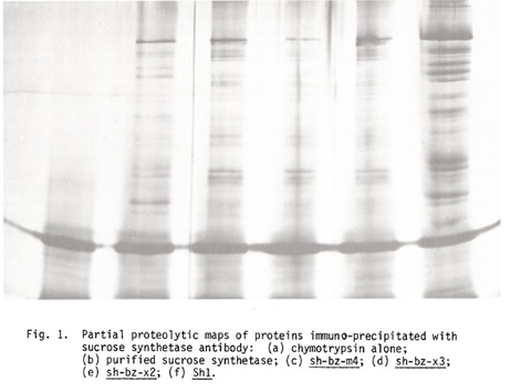
In order to obtain enough of the residual protein for peptide mapping, the sucrose synthetases were partially purified. 20 DAP endosperms of each strain were ground in 2 volumes of 10 mM Tris-HCl (pH 7.5), 10 mM MgCl2, and centrifuged at 23,000 g for 20 minutes. The supernatants were applied to Affigel-Blue columns equilibrated in the same buffer in the cold. The columns were washed with loading buffer and the sucrose synthetase fractions eluted at room temperature with 2 mM UMP, 10 mM NaCl and 10 mM Tris-HCl (pH 7.5). These concentrated fractions were immuno-precipitated in 0.15 M NaCl, 20 mM Tris-HCl (pH 7.5), 10% glycerol, 0.5% NP-40 and 0.5 mg/ml BSA. The antigen-antibody complexes were bound to protein A-Sepharose using 4 mg protein A-Sepharose/microliter of antiserum. The precipitates were washed twice with 0.5 M NaCl, 20 mM Tris-HCl (pH 7.5), 0.5% NP-40, and then once with the same buffer lacking NaCl. The antigen-antibody-protein A-Sepharose complexes were taken up in 20 mM DTT and 2% SDS, boiled for 2 minutes and applied to a 10% SDS acrylamide gel. This gel was stained for 30 minutes with Coomassie, destained one hour, and the 88K bands cut out. The gel pieces were inserted into the slots of a slightly wider SDS acrylamide gel and digested with chymotrypsin (7.5 micrograms/lane) as described by Cleveland et al. (J. Biol. Chem. 252:1102, 1977). This gel (Figure 1) was silver stained (Oakley et al., Anal. Biochem. 105:361, 1980).
It is evident that the 3 deletion strains (lanes c, d and e) have identical peptide map patterns that are different from the pattern in the Sh1 lanes (b and f). Chourey (1981) was able to detect the residual protein in a Sh1 extract by greatly overloading a native gel. There are probably peptide fragments from the residual sucrose synthetase that are beyond the level of detection in lanes b and f.
In summary, the peptide maps offer fairly conclusive evidence that the two sucrose synthetase proteins are coded by two different genes.
Sheila McCormick*
*Current address: IPRI, 853 Industrial Rd., San Carlos, CA 94403
Return to the MNL 56 On-Line Index
Return to the Maize Newsletter Index
Return to the MaizeGDB Homepage