One of the most powerful techniques for the separation of protein involves two dimensional resolution on polyacrylamide gel by molecular weight and charge. In situations where sample quantities are limited or speed of analysis is required, such separation methods are constrained. For these reasons, a variety of microgel systems have been developed (Ruchel, J. Histochem. Cytochem. 24:773-791, 1976; Condeelis, Anal. Biochem. 77:195-207, 1977) for the rapid reproducible analysis of nanogram quantities of protein. These micro-methods are almost exclusively used in the field of neurophysiology where the analysis of single large cells is desired.
It has long been observed that differences in Zea mays L. genotypes are expressed to varying degrees in the sporophytic generation. However, very few genotypic-phenotypic relationships are available in the gametophytic phase. Indeed, in the case of corn pollen, very few methods are capable of describing phenotypic variation among the grains from sporophytes of known backgrounds. Thus, new probes which can detect some components of genetic diversity in the gametophyte generation are needed urgently. A review of the biochemical literature revealed that approximately 100 enzymes have been identified in the tissues of the corn plant; fewer than 40 of these have been identified in the male and female gametophytes.
The electrophoretic separation of protein from plant tissue extracts is fundamentally the same as that employed in the resolution of animal tissue extracts. However, one primary difference resides in the method of protein extraction (see 6th contribution). The implementation and modification of a two dimensional microelectrophoretic technique originally reported by Ruchel (J. Chromat. 132:451-468, 1977) is described.
Hardware: A high voltage-low current power supply was built from a modified circuit outlined by Wurtzburg (Power Supply Handbook, Motorola Semiconductor Inc., Tucson Arizona, 1976). The power supply will function in either constant voltage (0-300 V) or constant current mode (0-100 µA) with less than .01% fluctuation in the electric field generated. As gel chambers, Drummond (Broomall, PA) 5 µl capillary tubes are employed in the first dimension and double diamond glass, plates, separated by teflon spacers, in the second. One of the glass plates is constructed with a rectangular notch (1 cm x 4 cm). The glass plates are fixed to the teflon spacers by a light coating of vacuum grease. The volume of the second dimension chamber is approximately 1000 µl (.08 x 4 x 3.5 cm) and has sufficient length to accommodate one cylindrical microgel (.07 x 3.3 cm). The first dimension buffer chamber is constructed by boring holes (1 to 6 usually) in the bottom of a 50 ml disposable plastic beaker. The rubber grommets supplied with the microcapillary tubes are inserted into the holes. Capillary gel tube chambers are inserted through the grommets thus forming the upper buffer reservoir. The lower reservoir is served by a 100 ml glass beaker on which the upper reservoir is seated. Platinum wire electrodes are fixed to both reservoirs. The second dimension buffer chamber is constructed of plexiglass to form a five-sided rectangular box. The long sides of the box are notched to match the notch on the second dimension slab gel chamber. Two gel chambers are clamped (with notches adjacent to each other) on opposite sides of the buffer chamber. A light coating of vacuum grease ensures a leakproof seal. This subassembly becomes the upper buffer chamber of the slab gel apparatus. The lower chamber, a large Coplin jar, allows sufficient area to insert the upper chamber. Platinum wire electrodes are fixed to both reservoirs.
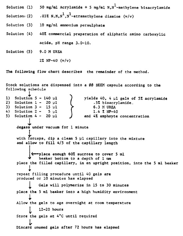
Preparation of isoelectric focussing (IEF) gels: The gel mixture has been modified from Ruchel (1976). The mixture is composed of stock solutions prepared fresh each week and stored in dark containers at 40 C. All solutions are prepared in double distilled water.
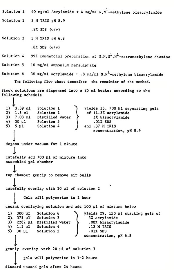
Preparation of sodium dodecyl sulphate (SDS) slab gels: The gel mixture employed in the second dimension has been modified from a method of Atkinson (Personal communication). The mixture is prepared by combining stock solutions prepared fresh each fortnight. All stock solutions are prepared in double distilled water and stored in dark containers at 40 C.
Loading and running IEF gels: Protein mixtures, stored on ice, are drawn up through a tuberculin syringe with a 30 gauge needle, then inserted into the upper end of the gel chamber by gentle pressure to the syringe. Failure to remove air bubbles in this procedure results in erratic conductivity during electrophoresis.
Microgels loaded with sample are inserted into the upper buffer chamber of the electrophoretic apparatus. The upper chamber is overlaid with anolyte (.5 N NaOH), the lower chamber with catholyte (.5 N H3PO4). The voltage is set to a constant field strength of 30 V/cm. Chromatophoric tracking dyes in the protein samples allow the monitoring of protein migration. Proteins are thought to be focused in the gel when the dye Xylene cyanole FF reaches the brownish precipitate of the faster migrating bromophenol blue (pI = 3.2).
Loading and running second dimension gels: The electrophoresed IEF gel is easily removed from the capillary by hydrostatic pressure delivered through a syringe fitted with 1/32" Tygon tubing. The gel is extruded into the notch of the second dimension gel chamber (previously attached to the upper buffer chamber), and pushed down onto the slab gel with a piece of filter paper. Immediately, running buffer (.1% SDS, .025 M TRIS pH 8.3, .2 M glycine, .001% Bromophenol Blue, .001% Xylene cyanole FF) is added to the upper buffer chamber. The lower buffer chamber is filled with running buffer (lacking tracking dye), and air bells are dispersed from the bottom and top of the gel chamber. A constant field strength of 100 µA/cm is allowed to flow from the top of the separation apparatus. The run is completed in approximately 30 minutes or when the marker dye just leaves the bottom of the gel.
Staining: The method outlined is employed as a general purpose staining procedure. All steps are performed at room temperature and with agitation of solutions: Remove gel from chamber; immerse in 10 volumes of fixer (10% glacial acetic acid + 50% methanol), prefix 15 minutes; transfer to staining solution (10 volumes of .1% Coomassie brilliant blue R-250 in 7% acetic acid + 30% methanol), stain 10 minutes; transfer to destain solution (10 volumes of 7% acetic acid + 10% methanol), destain 1 to 2 hours; transfer to fixer solution (10 volumes of 7% acetic acid + 5% methanol + 2% glycerol), postfix 15 minutes; photography; dehydration/storage.
The method will resolve nanogram quantities of protein. However, the differential dye binding capacity of some proteins will not allow this method to be employed in absolute protein quantitation. Alternate protein visualization methods are presently being tested.
Either dimension may be used separately. For separation on the basis of isoelectric point, cylindrical gels are immediately stained. Separation on the basis of molecular weight is accomplished by a slight modification to the slab gel apparatus. A template comb (cut from .8 mm teflon material) is inserted into solution 3 before polymerization takes place. Removal of the comb after polymerization leaves 8 wells (1 cm long x 3 mm wide) in the stacking gel of the slab. Gels produced in this way are attached to the buffer chamber apparatus as previously described. The assembled buffer chamber is then filled with running buffer and 1 to 5 µl of protein sample solutions are applied to each well. Electrophoresis and staining are carried out as before.
Employing the above microelectrophoretic procedures we have analyzed a number of tissues of the maize plant (e.g. pollen, primary root tips and leaf mesocotyl protoplasts). The results obtained with the micromethod are consistent with those yielded by the analysis of replicate samples employing a modified O'Farrel (J. Biol. Chem. 10:4007-4021, 1975) technique. However, the micromethod does allow a 100-fold increase in protein detection sensitivity by Coomassie brilliant blue R-250 staining, decreased preparation time, and smaller protein sample volumes for analysis.
W. G. Hughes
Return to the MNL 54 On-Line Index
Return to the Maize Newsletter Index
Return to the MaizeGDB Homepage