Eleven dominant disease lesion mutants were isolated in plantings of kernels from crosses by pollen treated with ethylmethane-sulfonate using the paraffin oil technique (Neuffer 1978). All eleven mutants were isolated as single plants in the M1 population. These plants were selfed and outcrossed to normal in succeeding generations using lesion mutants as a male parent. In the outcross population the segregation for mutant to normal was always found to be 1 mutant:1 normal indicating dominance.
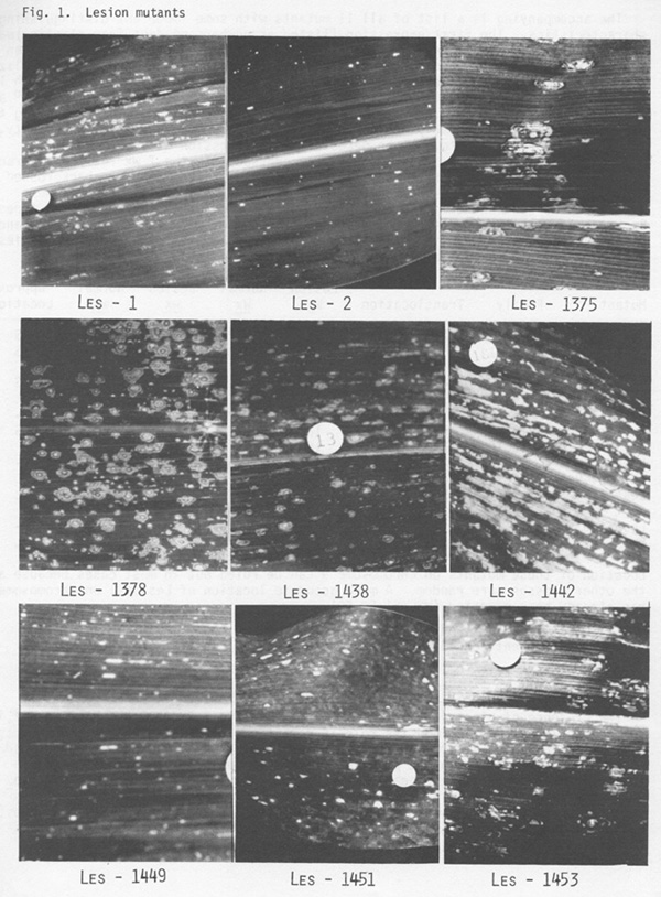
Characterization: The sequence of events that leads to lesion formation following infection of a susceptible host by a pathogen and the basic phenomena behind these events in plants have been extensively studied and are thought to be fairly well understood. In simplistic fashion we may say the pathogen produces a toxin which destroys the integrity of cell membranes and allows the successive invasion of surrounding cells. As a consequence spots with a center of dead cells and concentric rings of dying and newly interrupted cells occur on the leaves and other parts of the host plant. The frequency, size, shape, texture, color and intensity of lesions depends upon the genotypes of the host and the pathogen and in a different way upon the conditions at the time of infection. The disease lesion mutants reported by Neuffer and Calvert (J.H. 66:265-271, 1975) and those reported here produce a phenotype that mimics the symptoms of a particular disease under particular conditions. Sometimes the resemblance is so precise that the mimic cannot be distinguished from the disease except by testing for the presence or absence of the pathogen in the plant in question. Figure 1 presents sections of the 8th leaf of 45-day-old plants from 9 of the 11 mutants. Les*-1376 and Les*-1461 were not included because conditions were not ideal for their expression in the 1979 plantings used for photographing. Note differences in size, shape, intensity and distribution. For example Les1, Les*-1375 and Les*-1453 have large spreading lesions that closely resemble infection of susceptible lines of corn with several Helminthosporium species.
Les2, Les*-1442, Les*-1449 and Les*-1461 have small lesions; Les2 and
Les*-1449 resembling fungal infection of a "hypersensitive" resistant host
while Les*-1461 has small chlorotic spots resembling certain bacterial
diseases. Les*-1378 shows a distinctive clustering of both large and small
lesions.
| Mutant | Tissue Affected | First Expression | Temperature for Expression | Other Characteristics |
| Les1 | leaf, sheath | 10-12 | 20o C | - |
| Les2 | leaf | 12-20 | - | hypersensitive |
| Les*-1375 | leaf, sheath | 12-40 | - | large |
| Les*-1376 | leaf | 40 | - | like viral infection |
| Les*-1378 | leaf, sheath | 12-40 | - | random clusters |
| Les*-1438 | leaf, sheath | 12-20 | 200 C | - |
| Les*-1442 | leaf, sheath | 25-40 | - | follows vein; extreme necrosis |
| Les*-1449 | leaf | 25-40 | 320 C | hypersensitive |
| Les*-1451 | leaf, sheath | 12-40 | 200 C | chlorotic/necrotic spots |
| Les*-1453 | leaf, sheath | 12-40 | 200 C | large |
| Les*-1461 | leaf | - | - | chlorotic spots |
The accompanying is a list of all 11 mutants with some important distinguishing characteristics. The first expression (listed as numbers of days from planting) was taken under field conditions at Columbia, Missouri in June 1979; notes were taken at weekly intervals and individual plants varied a great deal, hence the generalized numbers. Temperature expression was obtained by growing in growth chambers with 14 hours moderate light and 10 hours of dark with constant temperatures of 200, 270 and 320 centigrade for 30 days. Those designated (-) did not express lesions during the experiment. This is probably due to the fact that other factors such as humidity, temperature variation, etc. may be necessary for expression of these mutants.
Location to chromosome: The mutants were crossed
with the T wx reciprocal translocation series available from the Coop.
Stock Center. The F1's were backcrossed to a homozygous wx non-mutant stock.
The resulting waxy and non-waxy kernels were separated and sown and the
resulting plants were noted for lesion and normal types. Backcross progenies
of most of the translocation stocks with the mutants gave random distribution
of wx and lesion characters. Following are those backcross progenies that
gave some indication of linkage:
| Backcross Classes | |||||||
| Mutant | Family | Translocation | Lesion Wx | Normal Wx | Lesion wx | Normal wx | Approx. Location |
| Les1 | 17:531 | 2-9b | 39 | 7 | 5 | 22 | 2S |
| Les2 | 27:2001 | 1-9c | 10 | 4 | 2 | 17 | 1S |
| 27:2002 | 13 | 7 | 3 | 16 | |||
| Les*-1375 | 27:2008 | 4-9g | 8 | 4 | 4 | 10 | 4S |
| Les*-1376 | 22:467 | 3-9c | 16 | 2 | 3 | 7 | 3L |
| Les*-1378 | - | - | - | - | - | - | - |
| Les*-1438 | 24:39 | 9-10b | 12 | 7 | 8 | 12 | 10S |
| 27:2016 | 16 | 9 | 11 | 21 | |||
| Les*-1442 | - | - | - | - | - | - | - |
| Les*-1449 | 27:2019 | 1-9(4997) | 15 | 4 | 5 | 15 | 1L |
| Les*-1451 | 26:48 | 5-9a | 22 | 17 | 15 | 22 | 5L |
| 27:2027 | 23 | 14 | 8 | 20 | |||
| Les*-1453 | - | - | - | - | - | - | - |
| Les*-1461 | 24:75 | 1-9(8389) | 16 | 4 | 4 | 15 | 1L |
Location of these mutants on chromosome 9 can be ruled out in most cases because all the other crosses were random. A more accurate location of Lesion-1 on chromosome 2 has already been reported (MNL 51:59-60).
M. G. Neuffer and S. E. Pawar*
*Permanent address: Bhabha Atomic Research Center, Trombay, Bombay, India
Return to the MNL 54 On-Line Index
Return to the Maize Newsletter Index
Return to the MaizeGDB Homepage